Download Mendeley
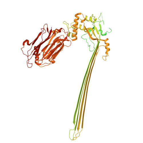
Molecular mechanism of pore formation by Plasmodium Perforin-like Protein 2.
Zhang, Y., Zhong, L., Song, Y., Guo, M., Ren, K., Yang, T., Huang, Y., Sirotkin, I., Yi, G., Jiao, F., Zhang, P., Gilbert, R.J.C., Ni, T., Yu, X.(2026) Nat Commun 17
- PubMed: 42675073 Search on PubMedSearch on PubMed Central
- DOI: https://doi.org/10.1038/s41467-026-76236-6
- Primary Citation Related Structures:
9XDA, 9XDB - PubMed Abstract:
Malaria-causing Plasmodium parasites must pass through several host cell types to complete their life cycle. This cell traversal is facilitated by perforin-like proteins (PLPs), among which PLP2 is essential for erythrocyte rupture by gametocytes. However, the mechanism by which PLP2 forms pores is not yet understood. Here, we combine cryo-electron microscopy and tomography to reveal the structural basis of Plasmodium vivax PLP2-mediated membrane attack. PvPLP2 assembles on lipid bilayers into heterogeneous arc- and ring-shaped pores with variable stoichiometries. Among them, we determine the structure of a 17-subunit pore complex in which the pore-forming MACPF domains form the central β-barrel, while the peripheral Apicomplexan PLP C-terminal β-pleated sheet (APCβ) domains anchor the complex to the membrane surface. A disulfide-stabilized mutant captures an intermediate pre-pore complex prior to membrane insertion, delineating the structural transitions that underpin β-barrel deployment. Functionally, PvPLP2 acts preferentially on the inner leaflet of the erythrocyte membrane, a specificity driven by its affinity for negatively charged lipids. Together, these findings establish the pore-formation pathway for a key Plasmodium virulence factor and provide a structural framework for rational design of transmission-blocking agents that prevent gametocyte egress.
- School of Biomedical Sciences, Li Ka Shing Faculty of Medicine, The University of Hong Kong, Hong Kong SAR, China.
Organizational Affiliation: