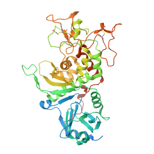
9TP2 | pdb_00009tp2

HomA ecto-domain mutant H350A E351A


Experimental Data Snapshot

  • Method: X-RAY DIFFRACTION
  • Resolution: 1.40 Å
  • R-Value Free: 
    0.230 (Depositor), 0.246 (DCC) 
  • R-Value Work: 
    0.209 (Depositor), 0.209 (DCC) 
  • R-Value Observed: 
    0.209 (Depositor) 

wwPDB Validation 3D Report Full Report

Validation slider image for 9TP2

This is version 1.1 of the entry. See complete history

Literature

Helicolysins Emerge as a New Family of Metzincin Metallopeptidases in Host-pathogen Interactions.

Rodriguez-Banqueri, A.Goulas, T.Girbal-Gonzalez, M.Vorderegger, S.Jarzab, M.Zarzecka, U.Guevara, T.Rodriguez-Lagunas, M.J.Eckhard, U.Perez-Cano, F.J.Wessler, S.Gomis-Ruth, F.X.

(2026) J Mol Biol 438: 169962-169962

  • DOI: https://doi.org/10.1016/j.jmb.2026.169962
  • Primary Citation Related Structures: 
    9TP2

  • PubMed Abstract: 

    The Helicobacter pylori Hom group of outer-membrane proteins (OMPs) has been implicated in virulence-related processes. Here, we present a structural and functional characterization of HomA from H. pylori strain 26695. Comparative in vivo analyses of the wild-type strain and a homA deletion mutant demonstrate a role for HomA in focal adhesion kinase phosphorylation at Y 397 and in bacterial adhesion to human leukaemia-derived MEC-1 cells. Recombinant production of several HomA variants identified a soluble ectodomain ( ECD HomA) that harbours the conserved zinc-binding motif of metzincin metallopeptidases (MPs) and displays dimerization capacity, acid resistance, and immunomodulatory activity by triggering a pro-inflammatory response in Raji human B-lymphoblastoid cancer cells. Site-directed mutagenesis of the zinc-binding motif confirmed its contribution to zinc coordination and thermal stability. Structural analysis of ECD HomA revealed a two-domain organization comprising an N-terminal accessory β-sandwich domain (ND) and a C-terminal catalytic domain (CD), in which a non-canonical 'Thr-turn' replaces the hallmark metzincin 'Met-turn'. Integrating bioinformatics with AI-based structure predictions uncovered putative HomA homologues across 44 proteomes, predominantly from Gram-negative bacteria. Structural and biochemical characterization of the closest non-Helicobacter homologue from the oral pathogen Capnocytophaga canimorsus, dubbed 'canilysin', revealed an architecture equivalent to ECD HomA and highly specific MP activity. Together, these findings define the Hom group and canilysin as helicolysins, a previously uncharacterized metzincin family distinguished by a conserved Thr-turn and an accessory ND, and implicates these proteins in host-pathogen interactions, adhesion, and immunomodulation.


  • Organizational Affiliation
    • Proteolysis Laboratory, Department of Structural Biology, Molecular Biology Institute of Barcelona (CSIC), Barcelona Science Park, c/Baldiri Reixac, 15-21, 08028 Barcelona, Catalonia, Spain. Electronic address: arbcri@ibmb.csic.es.

Macromolecule Content 

  • Total Structure Weight: 55.29 kDa 
  • Atom Count: 3,614 
  • Modeled Residue Count: 401 
  • Deposited Residue Count: 487 
  • Unique protein chains: 1

Macromolecules

Find similar proteins by:|  3D Structure
Entity ID: 1
MoleculeChains  Sequence LengthOrganismDetailsImage
HomA487Helicobacter pylori 26695Mutation(s): 2 
Gene Names: HP_0710
UniProt
Find proteins for O25414 (Helicobacter pylori (strain ATCC 700392 / 26695))
Explore O25414 
Go to UniProtKB:  O25414
Entity Groups
Sequence Clusters30% Identity50% Identity70% Identity90% Identity95% Identity100% Identity
UniProt GroupO25414
Sequence Annotations
Expand
Reference Sequence

Small Molecules

Ligands 4 Unique
IDChains Name / Formula / InChI Key2D Diagram3D Interactions
IOD

Query on IOD


Download:Ideal Coordinates CCD File
C [auth A]
D [auth A]
E [auth A]
F [auth A]
G [auth A]
C [auth A],
D [auth A],
E [auth A],
F [auth A],
G [auth A],
H [auth A],
I [auth A],
J [auth A]
IODIDE ION
I
XMBWDFGMSWQBCA-UHFFFAOYSA-M
EDO

Query on EDO


Download:Ideal Coordinates CCD File
B [auth A]1,2-ETHANEDIOL
C2 H6 O2
LYCAIKOWRPUZTN-UHFFFAOYSA-N
CL

Query on CL


Download:Ideal Coordinates CCD File
K [auth A]
L [auth A]
M [auth A]
N [auth A]
O [auth A]
K [auth A],
L [auth A],
M [auth A],
N [auth A],
O [auth A],
P [auth A]
CHLORIDE ION
Cl
VEXZGXHMUGYJMC-UHFFFAOYSA-M
NA

Query on NA


Download:Ideal Coordinates CCD File
Q [auth A]SODIUM ION
Na
FKNQFGJONOIPTF-UHFFFAOYSA-N

Experimental Data & Validation

Experimental Data

  • Method: X-RAY DIFFRACTION
  • Resolution: 1.40 Å
  • R-Value Free:  0.230 (Depositor), 0.246 (DCC) 
  • R-Value Work:  0.209 (Depositor), 0.209 (DCC) 
  • R-Value Observed: 0.209 (Depositor) 
Space Group: C 1 2 1
Unit Cell:
Length ( Å )Angle ( ˚ )
a = 141.12α = 90
b = 44.6β = 111.56
c = 68.92γ = 90
Software Package:
Software NamePurpose
MxCuBEdata collection
PHENIXrefinement
XDSdata reduction
XSCALEdata scaling
PHENIXphasing

Structure Validation

View Full Validation Report


Entry History 

& Funding Information

Deposition Data


Funding OrganizationLocationGrant Number
Not funded--

Revision History  (Full details and data files)

  • Version 1.0: 2026-08-19
    Type: Initial release
  • Version 1.1: 2026-09-09
    Changes: Database references