9SNV | pdb_00009snv

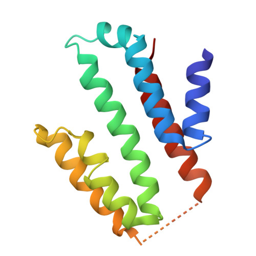
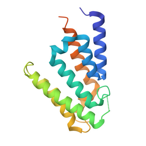
CTLH-CRA domains of the RanBP9-muskelin complex


Experimental Data Snapshot

  • Method: X-RAY DIFFRACTION
  • Resolution: 1.97 Å
  • R-Value Free: 
    0.255 (Depositor), 0.253 (DCC) 
  • R-Value Work: 
    0.228 (Depositor), 0.227 (DCC) 
  • R-Value Observed: 
    0.229 (Depositor) 

Starting Model: in silico
View more details

wwPDB Validation 3D Report Full Report

Validation slider image for 9SNV

This is version 1.1 of the entry. See complete history

Literature

A structural code for assembly specificity in GID/CTLH-type E3 ligases.

van gen Hassend, P.M.Schindelin, H.

(2026) Elife 15

  • DOI: https://doi.org/10.7554/eLife.110152
  • Primary Citation Related Structures: 
    9SNE, 9SNF, 9SNG, 9SNH, 9SNI, 9SNV, 9SOC, 9SOH, 9SOI

  • PubMed Abstract: 

    GID/CTLH-type E3 ligases assemble into conserved ring-shaped architectures built from repeating LisH-CTLH-CRA modules, yet the molecular rules that enforce their highly specific subunit arrangement have remained unknown. Here, we decode the structural 'assembly specificity code' that governs CRA-CRA pairing. Using crystal structures of multiple CTLH-CRA domains, including the RanBP9-muskelin heterodimer, integrated with quantitative binding analyses, we show that several interfaces operate with exceptionally high affinity, reaching the picomolar range, and that conserved sequence and geometric features enable each subunit to only select cognate partners. Strikingly, targeted perturbations of these features are sufficient to reprogram pairing preferences, enabling engineered subunits such as RanBP10 or Twa1 to adopt non-native interaction partners. These findings reveal the molecular logic that preserves the architecture of GID/CTLH-type E3 ligases and demonstrate that their assembly code is both decipherable and engineerable, providing a conceptual foundation for reconfiguring these ring-shaped E3 ligases.


  • Organizational Affiliation
    • Institute of Structural Biology, Rudolf Virchow Center for Integrative and Translational Bioimaging, Julius-Maximilians-Universität Würzburg, Würzburg, Germany.

Macromolecule Content 

  • Total Structure Weight: 35.32 kDa 
  • Atom Count: 2,229 
  • Modeled Residue Count: 272 
  • Deposited Residue Count: 309 
  • Unique protein chains: 2

Macromolecules

Find similar proteins by:|  3D Structure
Entity ID: 1
MoleculeChains  Sequence LengthOrganismDetailsImage
Muskelin137Rattus norvegicusMutation(s): 0 
Gene Names: Mkln1Msk
UniProt
Find proteins for Q99PV3 (Rattus norvegicus)
Explore Q99PV3 
Go to UniProtKB:  Q99PV3
Entity Groups
Sequence Clusters30% Identity50% Identity70% Identity90% Identity95% Identity100% Identity
UniProt GroupQ99PV3
Sequence Annotations
Expand
Reference Sequence
Find similar proteins by:|  3D Structure
Entity ID: 2
MoleculeChains  Sequence LengthOrganismDetailsImage
Ran-binding protein 9172Mus musculusMutation(s): 0 
Gene Names: Ranbp9Ranbpm
UniProt
Find proteins for P69566 (Mus musculus)
Explore P69566 
Go to UniProtKB:  P69566
Entity Groups
Sequence Clusters30% Identity50% Identity70% Identity90% Identity95% Identity100% Identity
UniProt GroupP69566
Sequence Annotations
Expand
Reference Sequence

Experimental Data & Validation

Experimental Data

  • Method: X-RAY DIFFRACTION
  • Resolution: 1.97 Å
  • R-Value Free:  0.255 (Depositor), 0.253 (DCC) 
  • R-Value Work:  0.228 (Depositor), 0.227 (DCC) 
  • R-Value Observed: 0.229 (Depositor) 
Space Group: P 61 2 2
Unit Cell:
Length ( Å )Angle ( ˚ )
a = 84.086α = 90
b = 84.086β = 90
c = 228.282γ = 120
Software Package:
Software NamePurpose
PHENIXrefinement
XDSdata reduction
STARANISOdata scaling
PHASERphasing

Structure Validation

View Full Validation Report


Entry History 

& Funding Information

Deposition Data


Funding OrganizationLocationGrant Number
German Research Foundation (DFG)GermanyGRK2243

Revision History  (Full details and data files)

  • Version 1.0: 2026-02-18
    Type: Initial release
  • Version 1.1: 2026-09-02
    Changes: Database references