9Q3L | pdb_00009q3l

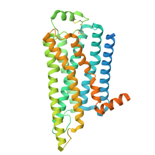
CryoEM structure of beta2-adrenergic receptor dimer mediated by a biased allosteric modulator in lipid nanodisc

  • Classification: SIGNALING PROTEIN
  • Organism(s): Homo sapiens
  • Expression System: Spodoptera frugiperda
  • Mutation(s): No 

  • Deposited: 2025-08-18 Released: 2026-08-19 
  • Deposition Author(s): Shen, J., Kobilka, B.K.
  • Funding Organization(s): National Institutes of Health/National Institute of Neurological Disorders and Stroke (NIH/NINDS), National Institutes of Health/National Institute of General Medical Sciences (NIH/NIGMS), American Heart Association

Experimental Data Snapshot

  • Method: ELECTRON MICROSCOPY
  • Resolution: 2.50 Å
  • Aggregation State: PARTICLE 
  • Reconstruction Method: SINGLE PARTICLE 

wwPDB Validation 3D Report Full Report

Validation slider image for 9Q3L

This is version 1.2 of the entry. See complete history

Literature

A biased allosteric modulator is a molecular glue for beta 2 AR dimerization.

Shen, J.Peddada, T.N.Komolov, K.E.De Pascali, F.Garces, A.M.Wang, H.Ehsan, M.Chae, P.S.Lerch, M.T.Benovic, J.L.Xu, J.Kobilka, B.K.

(2026) Nature 

  • DOI: https://doi.org/10.1038/s41586-026-10892-y
  • Primary Citation Related Structures: 
    9Q3L

  • PubMed Abstract: 

    Family A G-protein-coupled receptors (GPCRs) are typically described as monomers, yet growing evidence suggests that they can form dimers with distinct signalling properties 1-3 . However, the mechanisms and therapeutic potential of such dimerization remain poorly understood. Here we show that AP-7-168, an optimized derivative of a β-arrestin-biased negative allosteric modulator of the β 2 -adrenergic receptor (β 2 AR) that sustains bronchorelaxation in cell and tissue models 4 , functions as a molecular glue to stabilize β 2 AR homodimerization. Cryogenic electron microscopy structures reveal a unique binding mode in which two AP-7-168 molecules pack within a pocket formed by transmembrane helices 3, 4 and 5 of two protomers, stabilizing a dimeric conformation that selectively prevents β-arrestin coupling. In cells, AP-7-168 robustly stabilizes β 2 AR dimerization and drives enlarged nanocluster formation. Combined with extensive functional studies, our findings identify an allosteric mechanism by which a small molecule biases β 2 AR signalling through dimerization, highlighting ligand-stabilized dimerization as a strategy for GPCR modulation.


  • Organizational Affiliation
    • Department of Molecular and Cellular Physiology, Stanford University School of Medicine, Stanford, CA, USA.

Macromolecule Content 

  • Total Structure Weight: 106.1 kDa 
  • Atom Count: 4,790 
  • Modeled Residue Count: 574 
  • Deposited Residue Count: 920 
  • Unique protein chains: 1

Macromolecules

Find similar proteins by:|  3D Structure
Entity ID: 1
MoleculeChains  Sequence LengthOrganismDetailsImage
Beta-2 adrenergic receptor
A, B
460Homo sapiensMutation(s): 0 
Gene Names: ADRB2ADRB2RB2AR
UniProt & NIH Common Fund Data Resources
Find proteins for P07550 (Homo sapiens)
Explore P07550 
Go to UniProtKB:  P07550
PHAROS:  P07550
GTEx:  ENSG00000169252 
Entity Groups
Sequence Clusters30% Identity50% Identity70% Identity90% Identity95% Identity100% Identity
UniProt GroupP07550
Sequence Annotations
Expand
Reference Sequence

Small Molecules

Ligands 4 Unique
IDChains Name / Formula / InChI Key2D Diagram3D Interactions
A1CNS

Query on A1CNS


Download:Ideal Coordinates CCD File
F [auth A],
J [auth B]
6-bromo-N~4~-cyclohexyl-N~2~-(3,4-difluorophenyl)quinazoline-2,4-diamine
C20 H19 Br F2 N4
XPSOVAYWMLKDBS-UHFFFAOYSA-N
P0G

Query on P0G


Download:Ideal Coordinates CCD File
E [auth A],
I [auth B]
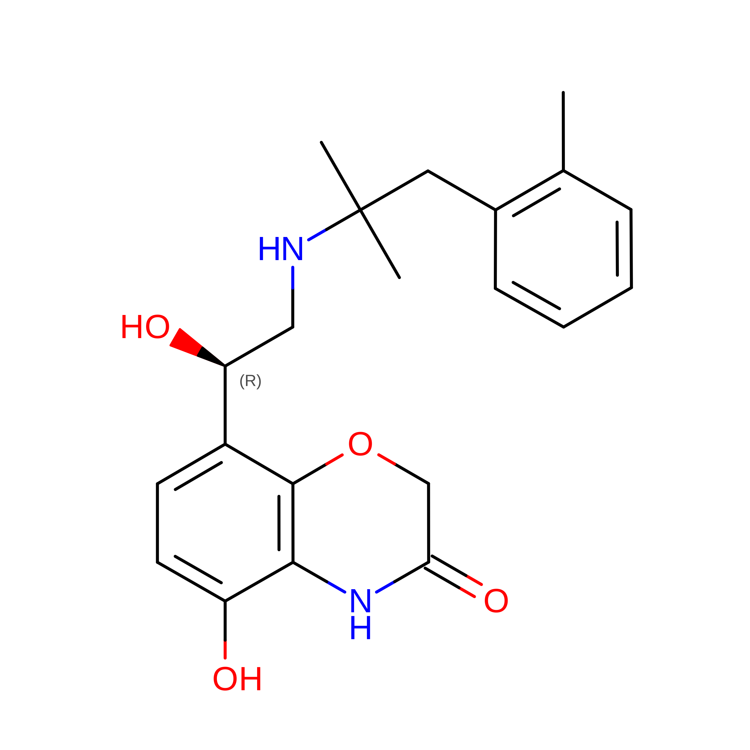
8-[(1R)-2-{[1,1-dimethyl-2-(2-methylphenyl)ethyl]amino}-1-hydroxyethyl]-5-hydroxy-2H-1,4-benzoxazin-3(4H)-one
C21 H26 N2 O4
NWQXBEWHTDRJIP-KRWDZBQOSA-N
LFA

Query on LFA


Download:Ideal Coordinates CCD File
D [auth A],
H [auth B]
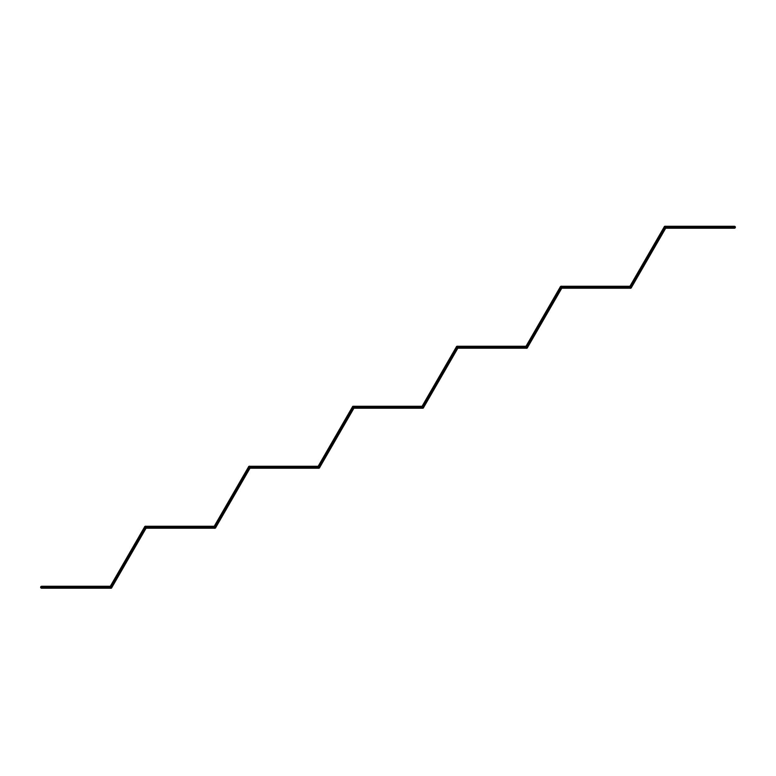
EICOSANE
C20 H42
CBFCDTFDPHXCNY-UHFFFAOYSA-N
C14

Query on C14


Download:Ideal Coordinates CCD File
C [auth A],
G [auth B]
TETRADECANE
C14 H30
BGHCVCJVXZWKCC-UHFFFAOYSA-N

Experimental Data & Validation

Experimental Data

  • Method: ELECTRON MICROSCOPY
  • Resolution: 2.50 Å
  • Aggregation State: PARTICLE 
  • Reconstruction Method: SINGLE PARTICLE 
EM Software:
TaskSoftware PackageVersion
RECONSTRUCTIONcryoSPARC

Structure Validation

View Full Validation Report


Entry History 

& Funding Information

Deposition Data


Funding OrganizationLocationGrant Number
National Institutes of Health/National Institute of Neurological Disorders and Stroke (NIH/NINDS)United StatesR35 NS137408
National Institutes of Health/National Institute of General Medical Sciences (NIH/NIGMS)United StatesR01 GM083118
American Heart AssociationUnited States25POST1411512

Revision History  (Full details and data files)

  • Version 1.0: 2026-08-19
    Type: Initial release
  • Version 1.1: 2026-08-26
    Changes: Data collection, Database references
  • Version 1.2: 2026-09-02
    Changes: Data collection, Database references