9N2K | pdb_00009n2k

N-terminal domain of Bacillus subtilis MutL bound to ADP


Experimental Data Snapshot

  • Method: X-RAY DIFFRACTION
  • Resolution: 1.36 Å
  • R-Value Free: 
    0.196 (Depositor), 0.196 (DCC) 
  • R-Value Work: 
    0.176 (Depositor), 0.177 (DCC) 
  • R-Value Observed: 
    0.176 (Depositor) 

Starting Model: experimental
View more details

wwPDB Validation   3D Report Full Report


Ligand Structure Quality Assessment 


This is version 1.1 of the entry. See complete history


Literature

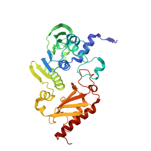
Bacillus subtilis MutL samples multiple conformations during nucleotide binding and hydrolysis.

Rodriguez Gonzalez, J.Davis, C.L.Wilkins, H.Erie, D.A.Guarne, A.

(2025) Structure 

  • DOI: https://doi.org/10.1016/j.str.2025.12.007
  • Primary Citation of Related Structures:  
    9N2K

  • PubMed Abstract: 

    DNA mismatch repair is an evolutionarily conserved repair pathway that corrects replication errors, thereby preventing genome instability. Two evolutionarily conserved proteins, MutS and MutL, recognize the mismatch and mark the newly synthesized strand for repair. Previous studies have shown how bacterial MutS homodimers function asymmetrically to recognize mismatches and recruit MutL. However, whether MutL homodimers also function asymmetrically to coordinate binding to MutS and activation of their nuclease activity remains unclear. Here, we characterize the ATPase domain of Bacillus subtilis MutL, a MutL protein with endonuclease activity, and delineate the differences with Escherichia coli MutL, a homolog without endonuclease activity. We find that B. subtilis MutL has low affinity for ATP and samples a repertoire of conformations that resemble those observed in eukaryotic MutL paralogs, indicating a relationship between ATP-induced dimer compaction and nuclease activity.


  • Organizational Affiliation
    • Department of Biochemistry and Centre de Recherche en Biologie Structurale, McGill University, Montreal, QC, Canada.

Macromolecules
Find similar proteins by:  (by identity cutoff)  |  3D Structure
Entity ID: 1
MoleculeChains Sequence LengthOrganismDetailsImage
DNA mismatch repair protein MutL
A, B
335Bacillus subtilisMutation(s): 0 
Gene Names: mutLBSU17050
UniProt
Find proteins for P49850 (Bacillus subtilis (strain 168))
Explore P49850 
Go to UniProtKB:  P49850
Entity Groups  
Sequence Clusters30% Identity50% Identity70% Identity90% Identity95% Identity100% Identity
UniProt GroupP49850
Sequence Annotations
Expand
  • Reference Sequence
Small Molecules
Ligands 3 Unique
IDChains Name / Formula / InChI Key2D Diagram3D Interactions
ADP (Subject of Investigation/LOI)
Query on ADP

Download Ideal Coordinates CCD File 
C [auth A],
I [auth B]
ADENOSINE-5'-DIPHOSPHATE
C10 H15 N5 O10 P2
XTWYTFMLZFPYCI-KQYNXXCUSA-N
SO4
Query on SO4

Download Ideal Coordinates CCD File 
E [auth A],
F [auth A],
G [auth A],
H [auth A],
K [auth B]
SULFATE ION
O4 S
QAOWNCQODCNURD-UHFFFAOYSA-L
MG (Subject of Investigation/LOI)
Query on MG

Download Ideal Coordinates CCD File 
D [auth A],
J [auth B]
MAGNESIUM ION
Mg
JLVVSXFLKOJNIY-UHFFFAOYSA-N
Experimental Data & Validation

Experimental Data

  • Method: X-RAY DIFFRACTION
  • Resolution: 1.36 Å
  • R-Value Free:  0.196 (Depositor), 0.196 (DCC) 
  • R-Value Work:  0.176 (Depositor), 0.177 (DCC) 
  • R-Value Observed: 0.176 (Depositor) 
Space Group: P 1 21 1
Unit Cell:
Length ( Å )Angle ( ˚ )
a = 56.72α = 90
b = 110.98β = 94.47
c = 57.41γ = 90
Software Package:
Software NamePurpose
PHENIXrefinement
MOSFLMdata reduction
pointlessdata scaling
PHENIXphasing

Structure Validation

View Full Validation Report


Ligand Structure Quality Assessment 


Entry History & Funding Information

Deposition Data


Funding OrganizationLocationGrant Number
Natural Sciences and Engineering Research Council (NSERC, Canada)Canada--

Revision History  (Full details and data files)

  • Version 1.0: 2026-01-07
    Type: Initial release
  • Version 1.1: 2026-01-14
    Changes: Database references