9N20 | pdb_00009n20

Structure of C3d Bound to a Fragment of FHR-2 and S. aureus Efb-C


Experimental Data Snapshot

  • Method: X-RAY DIFFRACTION
  • Resolution: 3.30 Å
  • R-Value Free: 
    0.214 (Depositor), 0.213 (DCC) 
  • R-Value Work: 
    0.178 (Depositor), 0.178 (DCC) 
  • R-Value Observed: 
    0.182 (Depositor) 

Starting Models: experimental
View more details

wwPDB Validation   3D Report Full Report


This is version 1.0 of the entry. See complete history


Literature

The C-terminal domain of Staphylococcus aureus Efb recruits FHR-2 to C3b, synergistically inhibiting the terminal complement pathway.

Duan, H.Kortvely, E.Mary, J.L.Hauck, S.M.Geisbrecht, B.V.

(2025) J Immunol 

  • DOI: https://doi.org/10.1093/jimmun/vkaf316
  • Primary Citation of Related Structures:  
    9N1Z, 9N20

  • PubMed Abstract: 

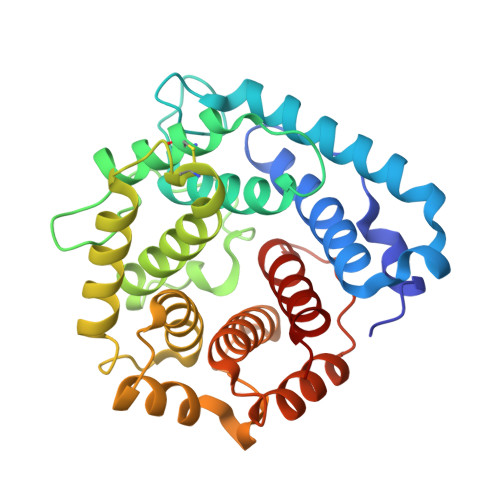
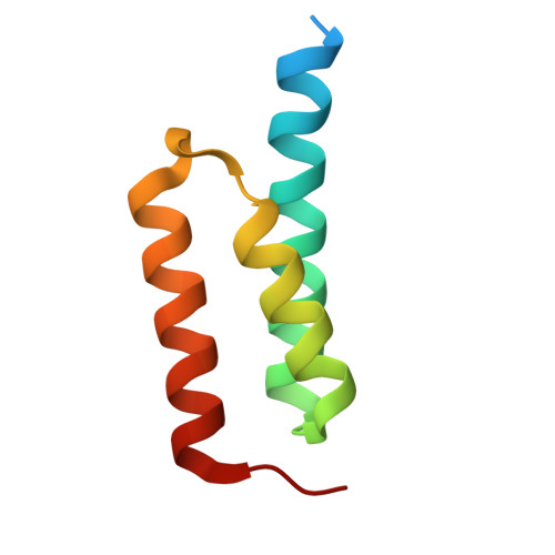
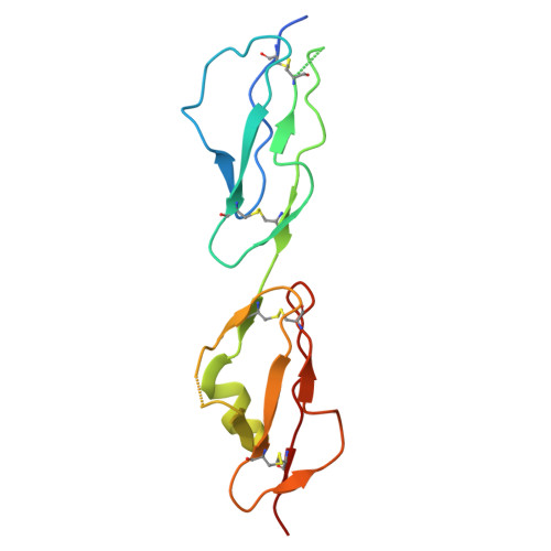
    The extracellular fibrinogen-binding protein (Efb) is one of nearly a dozen proteins secreted by Staphylococcus aureus to inhibit complement activation or amplification. The C-terminal domain of Efb (Efb-C) forms a high-affinity interaction with the thioester-containing domain of C3b (TED/C3d), thereby blocking formation of the C3 proconvertase complex through an allosteric mechanism. However, further functional consequences of Efb-C binding to C3b remain unexplored. Here, we identified a previously unknown interaction between Efb-C, C3b, and the complement regulatory molecule FHR2 (factor H-related protein 2). Since the FHR2/C3b interaction is centered upon the 2 C-terminal-most domains of FHR2 (FHR2[3-4]) and the TED/C3d domain of C3b, we tested whether Efb-C could influence the FHR2(3-4)/C3d interaction. We observed a significant enhancement of FHR2(3-4)/C3d binding in the presence of Efb-C. We studied the FHR2(3-4)/C3d/Efb-C complex by X-ray crystallography and found that Efb-C forms few direct interactions with FHR2(3-4). Yet, the presence of Efb-C also enhanced binding of FHR2(3-4) and full-length FHR2 to C3b, suggesting that the effect of Efb-C on the FHR2/C3b interaction arises from increased accessibility of the FHR2-binding site. We found that enhanced FHR2 binding did not impact the rate of C3 convertase formation more than Efb-C alone, nor did it impart decay acceleration or cofactor activity. However, we observed potent, synergistic inhibition of complement downstream of C5 activation by Efb-C and FHR2 but not by Efb-C and FHR2(3-4). Our results show that Efb-C binding to C3b exerts additional inhibitory effects on the central complement components beyond blocking formation of the C3 proconvertase alone.


  • Organizational Affiliation
    • Department of Biochemistry and Molecular Biophysics, Kansas State University, Manhattan, KS, United States.

Macromolecules
Find similar proteins by:  (by identity cutoff)  |  3D Structure
Entity ID: 1
MoleculeChains Sequence LengthOrganismDetailsImage
Complement C3dg fragment297Homo sapiensMutation(s): 1 
Gene Names: C3CPAMD1
UniProt & NIH Common Fund Data Resources
Find proteins for P01024 (Homo sapiens)
Explore P01024 
Go to UniProtKB:  P01024
PHAROS:  P01024
GTEx:  ENSG00000125730 
Entity Groups  
Sequence Clusters30% Identity50% Identity70% Identity90% Identity95% Identity100% Identity
UniProt GroupP01024
Sequence Annotations
Expand
  • Reference Sequence
Find similar proteins by:  (by identity cutoff)  |  3D Structure
Entity ID: 2
MoleculeChains Sequence LengthOrganismDetailsImage
Fibrinogen-binding proteinB [auth C]75Staphylococcus aureus subsp. aureus Mu50Mutation(s): 0 
Gene Names: fibefbSAV1158
UniProt
Find proteins for P68799 (Staphylococcus aureus (strain Mu50 / ATCC 700699))
Explore P68799 
Go to UniProtKB:  P68799
Entity Groups  
Sequence Clusters30% Identity50% Identity70% Identity90% Identity95% Identity100% Identity
UniProt GroupP68799
Sequence Annotations
Expand
  • Reference Sequence
Find similar proteins by:  (by identity cutoff)  |  3D Structure
Entity ID: 3
MoleculeChains Sequence LengthOrganismDetailsImage
Complement factor H-related protein 2C [auth B]131Homo sapiensMutation(s): 0 
Gene Names: CFHR2CFHL2FHR2HFL3
UniProt & NIH Common Fund Data Resources
Find proteins for P36980 (Homo sapiens)
Explore P36980 
Go to UniProtKB:  P36980
PHAROS:  P36980
GTEx:  ENSG00000080910 
Entity Groups  
Sequence Clusters30% Identity50% Identity70% Identity90% Identity95% Identity100% Identity
UniProt GroupP36980
Sequence Annotations
Expand
  • Reference Sequence
Experimental Data & Validation

Experimental Data

  • Method: X-RAY DIFFRACTION
  • Resolution: 3.30 Å
  • R-Value Free:  0.214 (Depositor), 0.213 (DCC) 
  • R-Value Work:  0.178 (Depositor), 0.178 (DCC) 
  • R-Value Observed: 0.182 (Depositor) 
Space Group: P 61
Unit Cell:
Length ( Å )Angle ( ˚ )
a = 145.584α = 90
b = 145.584β = 90
c = 76.855γ = 120
Software Package:
Software NamePurpose
PHENIXrefinement
HKL-2000data scaling
HKL-2000data reduction
PHASERphasing

Structure Validation

View Full Validation Report


Entry History & Funding Information

Deposition Data


Funding OrganizationLocationGrant Number
National Institutes of Health/National Institute of General Medical Sciences (NIH/NIGMS)United StatesR35GM140852

Revision History  (Full details and data files)

  • Version 1.0: 2025-12-10
    Type: Initial release