


Structure of nitrile synthetase ArtA
Ma, H.L., Zhang, K.K.To be published.
Experimental Data Snapshot
Starting Model: in silico
View more details
Entity ID: 1 | |||||
|---|---|---|---|---|---|
| Molecule | Chains | Sequence Length | Organism | Details | Image |
| Nitrile synthetase | 435 | Penicillium aurantiogriseum | Mutation(s): 0 | 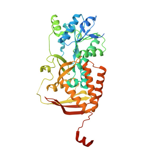 | |
Entity Groups | |||||
| Sequence Clusters | 30% Identity50% Identity70% Identity90% Identity95% Identity100% Identity | ||||
Sequence AnnotationsExpand | |||||
| |||||
| Ligands 1 Unique | |||||
|---|---|---|---|---|---|
| ID | Chains | Name / Formula / InChI Key | 2D Diagram | 3D Interactions | |
| GOL (Subject of Investigation/LOI) Query on GOL | E [auth A] F [auth A] G [auth B] H [auth B] I [auth C] | GLYCEROL C3 H8 O3 PEDCQBHIVMGVHV-UHFFFAOYSA-N |  | ||
| Length ( Å ) | Angle ( ˚ ) |
|---|---|
| a = 91.178 | α = 90 |
| b = 106.764 | β = 108.68 |
| c = 94.63 | γ = 90 |
| Software Name | Purpose |
|---|---|
| PHENIX | refinement |
| autoPX | data reduction |
| HKL-3000 | data scaling |
| PHENIX | phasing |
| Funding Organization | Location | Grant Number |
|---|---|---|
| National Natural Science Foundation of China (NSFC) | China | 32271358 |
| Other government | LZR202211060073 | |
| Other government | ZR2024QC065 |


