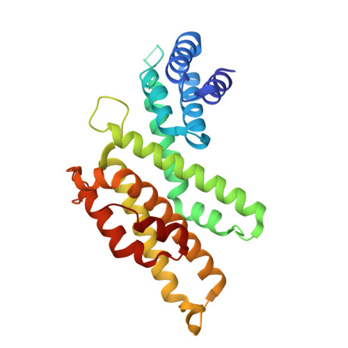
9E5A | pdb_00009e5a

Crystal structure of Acanthamoeba polyphaga mimivirus (APMV) vIF4G 53-312


Experimental Data Snapshot

  • Method: X-RAY DIFFRACTION
  • Resolution: 1.46 Å
  • R-Value Free: 
    0.196 (Depositor), 0.196 (DCC) 
  • R-Value Work: 
    0.176 (Depositor), 0.177 (DCC) 
  • R-Value Observed: 
    0.177 (Depositor) 

Starting Model: in silico
View more details

wwPDB Validation 3D Report Full Report

Validation slider image for 9E5A

This is version 1.1 of the entry. See complete history

Literature

Giant DNA viruses encode a hallmark translation initiation complex of eukaryotic life.

Fels, J.M.Hill, A.B.Han, R.Garcia, J.M.Bisio, H.Abergel, C.Kranzusch, P.J.Lee, A.S.Y.

(2026) Cell 189: 1423-1433.e16

  • DOI: https://doi.org/10.1016/j.cell.2026.01.008
  • Primary Citation Related Structures: 
    9E5A

  • PubMed Abstract: 

    In contrast to living organisms, viruses were long thought to lack protein synthesis machinery and instead depend on host factors to translate viral transcripts. Here, we discover that giant DNA viruses encode a distinct and functional IF4F translation-initiation complex to drive protein synthesis, thereby blurring the line between cellular and acellular biology. During infection, eukaryotic IF4F on host ribosomes is replaced by an essential viral IF4F that regulates viral translation, virion formation, and replication plasticity during altered host states. Structural dissection of viral IF4F reveals that the mRNA cap-binding subunit mediates exclusive interactions with viral mRNAs, constituting a molecular switch from translating host to viral proteins. Thus, our study establishes that viruses express a eukaryotic translation-initiation complex for protein synthesis, illuminating a series of evolutionary innovations in a core process of life.


  • Organizational Affiliation
    • Department of Cell Biology, Harvard Medical School, Boston, MA, USA; Department of Microbiology, Harvard Medical School, Boston, MA, USA; Department of Cancer Immunology and Virology, Dana-Farber Cancer Institute, Boston, MA, USA.

Macromolecule Content 

  • Total Structure Weight: 59.9 kDa 
  • Atom Count: 4,565 
  • Modeled Residue Count: 501 
  • Deposited Residue Count: 520 
  • Unique protein chains: 1

Macromolecules

Find similar proteins by:|  3D Structure
Entity ID: 1
MoleculeChains  Sequence LengthOrganismDetailsImage
Uncharacterized protein R255
A, B
260Acanthamoeba polyphaga mimivirusMutation(s): 0 
Gene Names: MIMI_R255
UniProt
Find proteins for Q5UPT9 (Acanthamoeba polyphaga mimivirus)
Explore Q5UPT9 
Go to UniProtKB:  Q5UPT9
Entity Groups
Sequence Clusters30% Identity50% Identity70% Identity90% Identity95% Identity100% Identity
UniProt GroupQ5UPT9
Sequence Annotations
Expand
Reference Sequence

Experimental Data & Validation

Experimental Data

  • Method: X-RAY DIFFRACTION
  • Resolution: 1.46 Å
  • R-Value Free:  0.196 (Depositor), 0.196 (DCC) 
  • R-Value Work:  0.176 (Depositor), 0.177 (DCC) 
  • R-Value Observed: 0.177 (Depositor) 
Space Group: P 1 21 1
Unit Cell:
Length ( Å )Angle ( ˚ )
a = 55.654α = 90
b = 91.317β = 117.54
c = 58.529γ = 90
Software Package:
Software NamePurpose
PHENIXrefinement
autoPROCdata reduction
autoPROCdata scaling
PHASERphasing

Structure Validation

View Full Validation Report


Entry History 

& Funding Information

Deposition Data


Funding OrganizationLocationGrant Number
National Institutes of Health/National Institute of General Medical Sciences (NIH/NIGMS)United States1DP2GM146250
National Institutes of Health/National Institute of General Medical Sciences (NIH/NIGMS)United States1R35GM142527

Revision History  (Full details and data files)

  • Version 1.0: 2026-02-25
    Type: Initial release
  • Version 1.1: 2026-09-09
    Changes: Database references