NMR Structure of Retinal Guanylate Cyclase Activating Protein 5 (GCAP5) with R22A Mutation That Abolishes Dimerization and Enhances Cyclase Activation.
Cudia, D.L., Ahoulou, E.O., Bej, A., Janssen, A.N., Scholten, A., Koch, K.W., Ames, J.B.(2024) Biochemistry 63: 1246-1256
- PubMed: 38662574
- DOI: https://doi.org/10.1021/acs.biochem.4c00046
- Primary Citation of Related Structures:
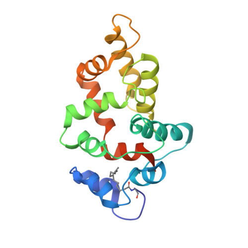
8VSX - PubMed Abstract:
Guanylate cyclase activating protein-5 (GCAP5) in zebrafish photoreceptors promotes the activation of membrane receptor retinal guanylate cyclase (GC-E). Previously, we showed the R22A mutation in GCAP5 (GCAP5 R22A ) abolishes dimerization of GCAP5 and activates GC-E by more than 3-fold compared to that of wild-type GCAP5 (GCAP5 WT ). Here, we present ITC, NMR, and functional analysis of GCAP5 R22A to understand how R22A causes a decreased dimerization affinity and increased cyclase activation. ITC experiments reveal GCAP5 R22A binds a total of 3 Ca 2+ , including two sites in the nanomolar range followed by a single micromolar site. The two nanomolar sites in GCAP5 WT were not detected by ITC, suggesting that R22A may affect the binding of Ca 2+ to these sites. The NMR-derived structure of GCAP5 R22A is overall similar to that of GCAP5 WT (RMSD = 2.3 Å), except for local differences near R22A (Q19, W20, Y21, and K23) and an altered orientation of the C-terminal helix near the N-terminal myristate. GCAP5 R22A lacks an intermolecular salt bridge between R22 and D71 that may explain the weakened dimerization. We present a structural model of GCAP5 bound to GC-E in which the R22 side-chain contacts exposed hydrophobic residues in GC-E. Cyclase assays suggest that GC-E binds to GCAP5 R22A with ∼25% higher affinity compared to GCAP5 WT , consistent with more favorable hydrophobic contact by R22A that may help explain the increased cyclase activation.
- Department of Chemistry, University of California, Davis, Davis, California 95616, United States.
Organizational Affiliation: