7SV2

Human Cytochrome P450 (CYP) 3A5 ternary complex with azamulin


Experimental Data Snapshot

  • Method: X-RAY DIFFRACTION
  • Resolution: 2.46 Å
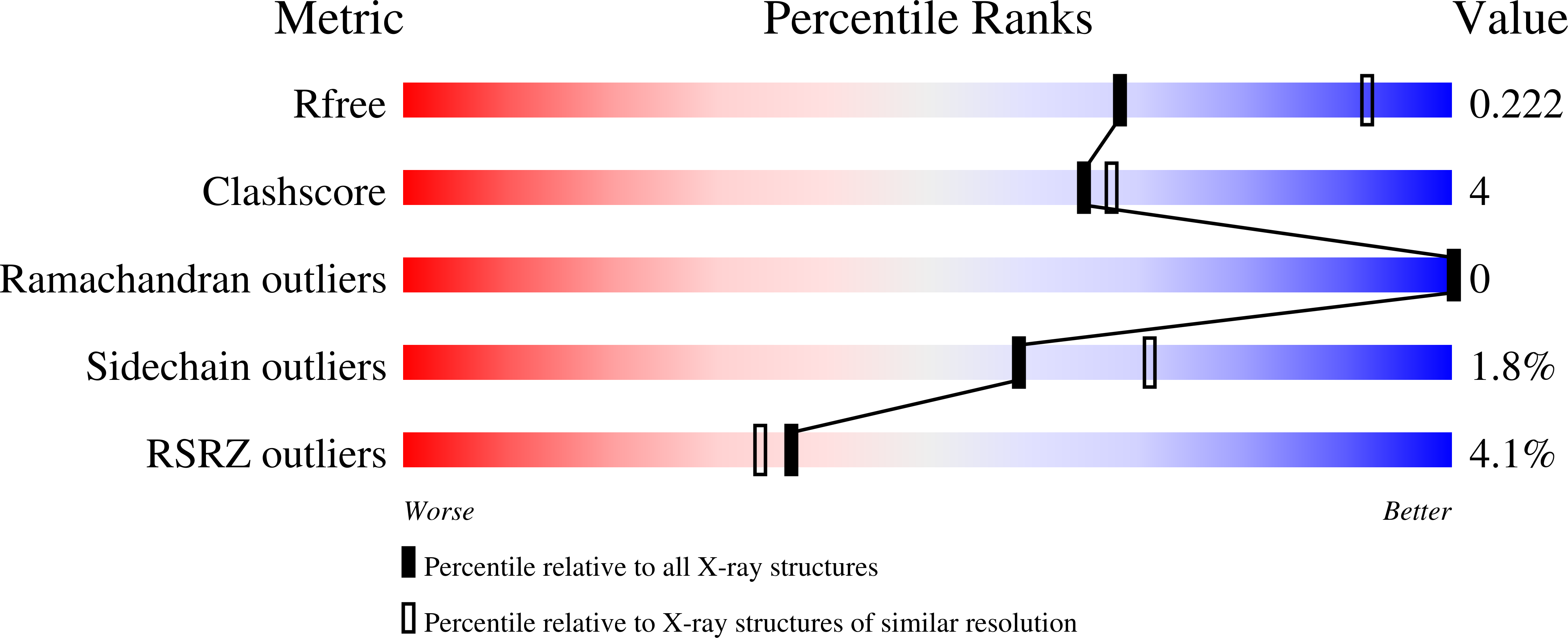
  • R-Value Free: 0.223 
  • R-Value Work: 0.188 
  • R-Value Observed: 0.189 

wwPDB Validation   3D Report Full Report


Ligand Structure Quality Assessment 


This is version 1.1 of the entry. See complete history


Literature

Structural characterization of the homotropic cooperative binding of azamulin to human cytochrome P450 3A5.

Hsu, M.H.Johnson, E.F.

(2022) J Biol Chem 298: 101909-101909

  • DOI: https://doi.org/10.1016/j.jbc.2022.101909
  • Primary Citation of Related Structures:  
    7SV2

  • PubMed Abstract: 

    Cytochrome P450 3A4 and 3A5 catalyze the metabolic clearance of a large portion of therapeutic drugs. Azamulin is used as a selective inhibitor for 3A4 and 3A5 to define their roles in metabolism of new chemical entities during drug development. In contrast to 3A4, 3A5 exhibits homotropic cooperativity for the sequential binding of two azamulin molecules at concentrations used for inhibition. To define the underlying sites and mechanisms for cooperativity, an X-ray crystal structure of 3A5 was determined with two azamulin molecules in the active site that are stacked in an antiparallel orientation. One azamulin resides proximal to the heme in a pose similar to the 3A4-azamulin complex. Comparison to the 3A5 apo structure indicates that the distal azamulin in 3A5 ternary complex causes a significant induced fit that excludes water from the hydrophobic surfaces of binding cavity and the distal azamulin, which is augmented by the stacking interaction with the proximal azamulin. Homotropic cooperativity was not observed for the binding of related pleuromutilin antibiotics, tiamulin, retapamulin, and lefamulin, to 3A5, which are larger and unlikely to bind in the distal site in a stacked orientation. Formation of the 3A5 complex with two azamulin molecules may prevent time-dependent inhibition that is seen for 3A4 by restricting alternate product formation and/or access of reactive intermediates to vulnerable protein sites. These results also contribute to a better understanding of sites for cooperative binding and the differential structural plasticity of 3A5 and 3A4 that contribute to differential substrate and inhibitor binding.


  • Organizational Affiliation

    Department of Molecular Medicine, Scripps Research, La Jolla, California, USA.


Macromolecules
Find similar proteins by:  (by identity cutoff)  |  3D Structure
Entity ID: 1
MoleculeChains Sequence LengthOrganismDetailsImage
Cytochrome P450 3A5
A, B, C, D
480Homo sapiensMutation(s): 0 
Gene Names: CYP3A5
EC: 1.14.14.1
Membrane Entity: Yes 
UniProt & NIH Common Fund Data Resources
Find proteins for P20815 (Homo sapiens)
Explore P20815 
Go to UniProtKB:  P20815
PHAROS:  P20815
GTEx:  ENSG00000106258 
Entity Groups  
Sequence Clusters30% Identity50% Identity70% Identity90% Identity95% Identity100% Identity
UniProt GroupP20815
Sequence Annotations
Expand
  • Reference Sequence
Small Molecules
Ligands 2 Unique
IDChains Name / Formula / InChI Key2D Diagram3D Interactions
HEM (Subject of Investigation/LOI)
Query on HEM

Download Ideal Coordinates CCD File 
E [auth A],
H [auth B],
K [auth C],
N [auth D]
PROTOPORPHYRIN IX CONTAINING FE
C34 H32 Fe N4 O4
KABFMIBPWCXCRK-RGGAHWMASA-L
MWY
Query on MWY

Download Ideal Coordinates CCD File 
F [auth A]
G [auth A]
I [auth B]
J [auth B]
L [auth C]
F [auth A],
G [auth A],
I [auth B],
J [auth B],
L [auth C],
M [auth C],
O [auth D],
P [auth D]
(3aS,4R,5S,6R,8R,9R,9aR,10R)-6-ethyl-5-hydroxy-4,6,9,10-tetramethyl-1-oxodecahydro-3a,9-propanocyclopenta[8]annulen-8-yl [(5-amino-1H-1,2,4-triazol-3-yl)sulfanyl]acetate
C24 H38 N4 O4 S
FMHQJXGMLMSMLC-WBUYAQKGSA-N
Experimental Data & Validation

Experimental Data

  • Method: X-RAY DIFFRACTION
  • Resolution: 2.46 Å
  • R-Value Free: 0.223 
  • R-Value Work: 0.188 
  • R-Value Observed: 0.189 
  • Space Group: C 2 2 21
Unit Cell:
Length ( Å )Angle ( ˚ )
a = 104.786α = 90
b = 134.3β = 90
c = 275.274γ = 90
Software Package:
Software NamePurpose
JBluIce-EPICSdata collection
PHENIXrefinement
XDSdata reduction
Aimlessdata scaling
PHASERphasing

Structure Validation

View Full Validation Report


Ligand Structure Quality Assessment 


Entry History & Funding Information

Deposition Data


Funding OrganizationLocationGrant Number
National Institutes of Health/National Institute of General Medical Sciences (NIH/NIGMS)United StatesGM031001

Revision History  (Full details and data files)

  • Version 1.0: 2022-05-18
    Type: Initial release
  • Version 1.1: 2023-10-18
    Changes: Data collection, Refinement description