7STO

Chitin Synthase 2 from Candida albicans bound to polyoxin D


Experimental Data Snapshot

  • Method: ELECTRON MICROSCOPY
  • Resolution: 3.15 Å
  • Aggregation State: PARTICLE 
  • Reconstruction Method: SINGLE PARTICLE 

wwPDB Validation   3D Report Full Report


This is version 1.1 of the entry. See complete history


Literature

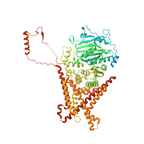
Structural basis for inhibition and regulation of a chitin synthase from Candida albicans.

Ren, Z.Chhetri, A.Guan, Z.Suo, Y.Yokoyama, K.Lee, S.Y.

(2022) Nat Struct Mol Biol 29: 653-664

  • DOI: https://doi.org/10.1038/s41594-022-00791-x
  • Primary Citation of Related Structures:  
    7STL, 7STM, 7STN, 7STO

  • PubMed Abstract: 

    Chitin is an essential component of the fungal cell wall. Chitin synthases (Chss) catalyze chitin formation and translocation across the membrane and are targets of antifungal agents, including nikkomycin Z and polyoxin D. Lack of structural insights into the action of these inhibitors on Chs has hampered their further development to the clinic. We present the cryo-EM structures of Chs2 from Candida albicans (CaChs2) in the apo, substrate-bound, nikkomycin Z-bound, and polyoxin D-bound states. CaChs2 adopts a unique domain-swapped dimer configuration where a conserved motif in the domain-swapped region controls enzyme activity. CaChs2 has a dual regulation mechanism where the chitin translocation tunnel is closed by the extracellular gate and plugged by a lipid molecule in the apo state to prevent non-specific leak. Analyses of substrate and inhibitor binding provide insights into the chemical logic of Chs inhibition, which can guide Chs-targeted antifungal development.


  • Organizational Affiliation

    Department of Biochemistry, Duke University School of Medicine, Durham, NC, USA.


Macromolecules
Find similar proteins by:  (by identity cutoff)  |  3D Structure
Entity ID: 1
MoleculeChains Sequence LengthOrganismDetailsImage
Chitin synthaseA [auth B],
B [auth A]
1,039Candida albicansMutation(s): 0 
Gene Names: CHS2CAALFM_CR09020CAorf19.7298
EC: 2.4.1.16
Membrane Entity: Yes 
UniProt
Find proteins for P30572 (Candida albicans)
Explore P30572 
Go to UniProtKB:  P30572
Entity Groups  
Sequence Clusters30% Identity50% Identity70% Identity90% Identity95% Identity100% Identity
UniProt GroupP30572
Sequence Annotations
Expand
  • Reference Sequence
Small Molecules
Ligands 2 Unique
IDChains Name / Formula / InChI Key2D Diagram3D Interactions
3PE (Subject of Investigation/LOI)
Query on 3PE

Download Ideal Coordinates CCD File 
C [auth B]
D [auth B]
E [auth B]
G [auth A]
H [auth A]
C [auth B],
D [auth B],
E [auth B],
G [auth A],
H [auth A],
I [auth A],
J [auth A],
L [auth A]
1,2-Distearoyl-sn-glycerophosphoethanolamine
C41 H82 N O8 P
LVNGJLRDBYCPGB-LDLOPFEMSA-N
BJ9 (Subject of Investigation/LOI)
Query on BJ9

Download Ideal Coordinates CCD File 
F [auth B],
K [auth A]
1-{(2R,3R,4S,5R)-5-[(S)-{[(2S,3S,4S)-2-amino-5-(carbamoyloxy)-3,4-dihydroxypentanoyl]amino}(carboxy)methyl]-3,4-dihydroxyoxolan-2-yl}-2,4-dioxo-1,2,3,4-tetrahydropyrimidine-5-carboxylic acid (non-preferred name)
C17 H23 N5 O14
JPFWJDMDPLEUBD-ITJAGOAWSA-N
Experimental Data & Validation

Experimental Data

  • Method: ELECTRON MICROSCOPY
  • Resolution: 3.15 Å
  • Aggregation State: PARTICLE 
  • Reconstruction Method: SINGLE PARTICLE 

Structure Validation

View Full Validation Report


Entry History & Funding Information

Deposition Data


Funding OrganizationLocationGrant Number
National Institutes of Health/National Institute of General Medical Sciences (NIH/NIGMS)R01GM115729

Revision History  (Full details and data files)

  • Version 1.0: 2022-07-13
    Type: Initial release
  • Version 1.1: 2022-07-27
    Changes: Database references