6WS1

Crystal structure of human phenylethanolamine N-methyltransferase (PNMT) in complex with (2S)-2-amino-4-((((2R,5R)-5-(6-amino-9H-purin-9-yl)-3,4-dihydroxytetrahydrofuran-2-yl)methyl)(3-(7,8-dichloro-1,2,3,4-tetrahydroisoquinolin-4-yl)propyl)amino)butanoic acid and AdoHcy (SAH)


Experimental Data Snapshot

  • Method: X-RAY DIFFRACTION
  • Resolution: 2.76 Å
  • R-Value Free: 0.274 
  • R-Value Work: 0.226 
  • R-Value Observed: 0.229 

wwPDB Validation   3D Report Full Report


Ligand Structure Quality Assessment 


This is version 1.2 of the entry. See complete history


Literature

Transition-State Analogues of PhenylethanolamineN-Methyltransferase.

Mahmoodi, N.Harijan, R.K.Schramm, V.L.

(2020) J Am Chem Soc 142: 14222-14233

  • DOI: https://doi.org/10.1021/jacs.0c05446
  • Primary Citation of Related Structures:  
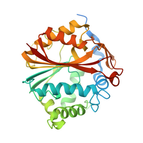
    6WS1

  • PubMed Abstract: 

    Phenylethanolamine N -methyltransferase (PNMT) is a critical enzyme in catecholamine synthesis. It transfers the methyl group of S -adenosylmethionine (SAM) to catalyze the synthesis of epinephrine from norepinephrine. Epinephrine has been associated with diverse human processes, including the regulation of blood pressure and respiration, as well as neurodegeneration found in Alzheimer's disease. Human PNMT (hPNMT) proceeds through an S N 2 transition state (TS) in which the transfer of the methyl group is rate limiting. TS analogue enzyme inhibitors are specific for their target and bind orders of magnitude more tightly than their substrates. Molecules resembling the TS of hPNMT were designed, synthesized, and kinetically characterized. This new inhibitory scaffold was designed to mimic the geometry and electronic properties of the hPNMT TS. Synthetic efforts resulted in a tight-binding inhibitor with a K i value of 12.0 nM. This is among the first of the TS analogue inhibitors of methyltransferase enzymes to show an affinity in the nanomolar range. Isothermal titration calorimetry (ITC) measurements indicated negative cooperative binding of inhibitor to the dimeric protein, driven by favorable entropic contributions. Structural analysis revealed that inhibitor 3 binds to hPNMT by filling the catalytic binding pockets for the cofactor (SAM) and the substrate (norepinephrine) binding sites.


  • Organizational Affiliation

    Department of Biochemistry, Albert Einstein College of Medicine, New York, New York 10461, United States.


Macromolecules
Find similar proteins by:  (by identity cutoff)  |  3D Structure
Entity ID: 1
MoleculeChains Sequence LengthOrganismDetailsImage
Phenylethanolamine N-methyltransferase
A, B
294Homo sapiensMutation(s): 0 
Gene Names: PNMTPENT
EC: 2.1.1.28
UniProt & NIH Common Fund Data Resources
Find proteins for P11086 (Homo sapiens)
Explore P11086 
Go to UniProtKB:  P11086
PHAROS:  P11086
GTEx:  ENSG00000141744 
Entity Groups  
Sequence Clusters30% Identity50% Identity70% Identity90% Identity95% Identity100% Identity
UniProt GroupP11086
Sequence Annotations
Expand
  • Reference Sequence
Small Molecules
Ligands 5 Unique
IDChains Name / Formula / InChI Key2D Diagram3D Interactions
U87 (Subject of Investigation/LOI)
Query on U87

Download Ideal Coordinates CCD File 
H [auth B]5'-([(3S)-3-amino-3-carboxypropyl]{3-[(4R)-7,8-dichloro-1,2,3,4-tetrahydroisoquinolin-4-yl]propyl}amino)-5'-deoxyadenosine
C26 H34 Cl2 N8 O5
LJUSEDZPCVZRMF-RYHYOYCJSA-N
SAH (Subject of Investigation/LOI)
Query on SAH

Download Ideal Coordinates CCD File 
C [auth A]S-ADENOSYL-L-HOMOCYSTEINE
C14 H20 N6 O5 S
ZJUKTBDSGOFHSH-WFMPWKQPSA-N
CD (Subject of Investigation/LOI)
Query on CD

Download Ideal Coordinates CCD File 
F [auth A]CADMIUM ION
Cd
WLZRMCYVCSSEQC-UHFFFAOYSA-N
EDO (Subject of Investigation/LOI)
Query on EDO

Download Ideal Coordinates CCD File 
D [auth A],
E [auth A],
I [auth B]
1,2-ETHANEDIOL
C2 H6 O2
LYCAIKOWRPUZTN-UHFFFAOYSA-N
K (Subject of Investigation/LOI)
Query on K

Download Ideal Coordinates CCD File 
G [auth A]POTASSIUM ION
K
NPYPAHLBTDXSSS-UHFFFAOYSA-N
Experimental Data & Validation

Experimental Data

  • Method: X-RAY DIFFRACTION
  • Resolution: 2.76 Å
  • R-Value Free: 0.274 
  • R-Value Work: 0.226 
  • R-Value Observed: 0.229 
  • Space Group: P 43 21 2
Unit Cell:
Length ( Å )Angle ( ˚ )
a = 93.886α = 90
b = 93.886β = 90
c = 188.272γ = 90
Software Package:
Software NamePurpose
PHENIXrefinement
PDB_EXTRACTdata extraction
iMOSFLMdata reduction
Aimlessdata scaling
PHASERphasing

Structure Validation

View Full Validation Report


Ligand Structure Quality Assessment 


Entry History & Funding Information

Deposition Data


Funding OrganizationLocationGrant Number
National Institutes of Health/National Institute of General Medical Sciences (NIH/NIGMS)United States--

Revision History  (Full details and data files)

  • Version 1.0: 2020-08-05
    Type: Initial release
  • Version 1.1: 2020-09-02
    Changes: Database references
  • Version 1.2: 2023-10-18
    Changes: Advisory, Data collection, Database references, Refinement description