6SY1

Crystal structure of the human 2-oxoadipate dehydrogenase DHTKD1 (E1)


Experimental Data Snapshot

  • Method: X-RAY DIFFRACTION
  • Resolution: 1.87 Å
  • R-Value Free: 0.230 
  • R-Value Work: 0.190 
  • R-Value Observed: 0.192 

wwPDB Validation   3D Report Full Report


Ligand Structure Quality Assessment 


This is version 1.4 of the entry. See complete history


Literature

Crystal structure and interaction studies of human DHTKD1 provide insight into a mitochondrial megacomplex in lysine catabolism.

Bezerra, G.A.Foster, W.R.Bailey, H.J.Hicks, K.G.Sauer, S.W.Dimitrov, B.McCorvie, T.J.Okun, J.G.Rutter, J.Kolker, S.Yue, W.W.

(2020) IUCrJ 7: 693-706

  • DOI: https://doi.org/10.1107/S205225252000696X
  • Primary Citation of Related Structures:  
    6SY1

  • PubMed Abstract: 

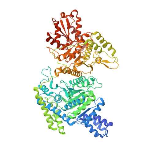
    DHTKD1 is a lesser-studied E1 enzyme among the family of 2-oxoacid de-hydrogenases. In complex with E2 (di-hydro-lipo-amide succinyltransferase, DLST) and E3 (dihydrolipo-amide de-hydrogenase, DLD) components, DHTKD1 is involved in lysine and tryptophan catabolism by catalysing the oxidative de-carboxyl-ation of 2-oxoadipate (2OA) in mitochondria. Here, the 1.9 Å resolution crystal structure of human DHTKD1 is solved in complex with the thi-amine diphosphate co-factor. The structure reveals how the DHTKD1 active site is modelled upon the well characterized homologue 2-oxoglutarate (2OG) de-hydrogenase but engineered specifically to accommodate its preference for the longer substrate of 2OA over 2OG. A 4.7 Å resolution reconstruction of the human DLST catalytic core is also generated by single-particle electron microscopy, revealing a 24-mer cubic scaffold for assembling DHTKD1 and DLD protomers into a megacomplex. It is further demonstrated that missense DHTKD1 variants causing the inborn error of 2-amino-adipic and 2-oxoadipic aciduria impact on the complex formation, either directly by disrupting the interaction with DLST, or indirectly through destabilizing the DHTKD1 protein. This study provides the starting framework for developing DHTKD1 modulators to probe the intricate mitochondrial energy metabolism.


  • Organizational Affiliation

    Structural Genomics Consortium, Nuffield Department of Medicine, University of Oxford, Oxford, OX3 7DQ, United Kingdom.


Macromolecules
Find similar proteins by:  (by identity cutoff)  |  3D Structure
Entity ID: 1
MoleculeChains Sequence LengthOrganismDetailsImage
Probable 2-oxoglutarate dehydrogenase E1 component DHKTD1, mitochondrial
A, B
898Homo sapiensMutation(s): 0 
Gene Names: DHTKD1KIAA1630
EC: 1.2.4.2
UniProt & NIH Common Fund Data Resources
Find proteins for Q96HY7 (Homo sapiens)
Explore Q96HY7 
Go to UniProtKB:  Q96HY7
PHAROS:  Q96HY7
GTEx:  ENSG00000181192 
Entity Groups  
Sequence Clusters30% Identity50% Identity70% Identity90% Identity95% Identity100% Identity
UniProt GroupQ96HY7
Sequence Annotations
Expand
  • Reference Sequence
Experimental Data & Validation

Experimental Data

  • Method: X-RAY DIFFRACTION
  • Resolution: 1.87 Å
  • R-Value Free: 0.230 
  • R-Value Work: 0.190 
  • R-Value Observed: 0.192 
  • Space Group: P 1
Unit Cell:
Length ( Å )Angle ( ˚ )
a = 78.55α = 63.43
b = 81.22β = 76.96
c = 86.9γ = 72.06
Software Package:
Software NamePurpose
xia2data scaling
PHENIXrefinement
PDB_EXTRACTdata extraction
xia2data reduction
PHASERphasing

Structure Validation

View Full Validation Report


Ligand Structure Quality Assessment 


Entry History & Funding Information

Deposition Data


Funding OrganizationLocationGrant Number
Wellcome TrustUnited Kingdom092809/Z/10/Z

Revision History  (Full details and data files)

  • Version 1.0: 2020-06-24
    Type: Initial release
  • Version 1.1: 2020-10-21
    Changes: Database references, Derived calculations
  • Version 1.2: 2020-10-28
    Changes: Structure summary
  • Version 1.3: 2021-08-04
    Changes: Data collection
  • Version 1.4: 2024-01-24
    Changes: Data collection, Database references, Refinement description