6SIT

Pseudo-atomic crystal structure of the desmoglein 2 - human adenovirus serotype 3 fibre knob complex


Experimental Data Snapshot

  • Method: X-RAY DIFFRACTION
  • Resolution: 4.50 Å
  • R-Value Free: 0.370 
  • R-Value Work: 0.367 

wwPDB Validation   3D Report Full Report


This is version 1.1 of the entry. See complete history


Literature

Intermediate-resolution crystal structure of the human adenovirus B serotype 3 fibre knob in complex with the EC2-EC3 fragment of desmoglein 2.

Vassal-Stermann, E.Hutin, S.Fender, P.Burmeister, W.P.

(2019) Acta Crystallogr F Struct Biol Commun 75: 750-757

  • DOI: https://doi.org/10.1107/S2053230X19015784
  • Primary Citation of Related Structures:  
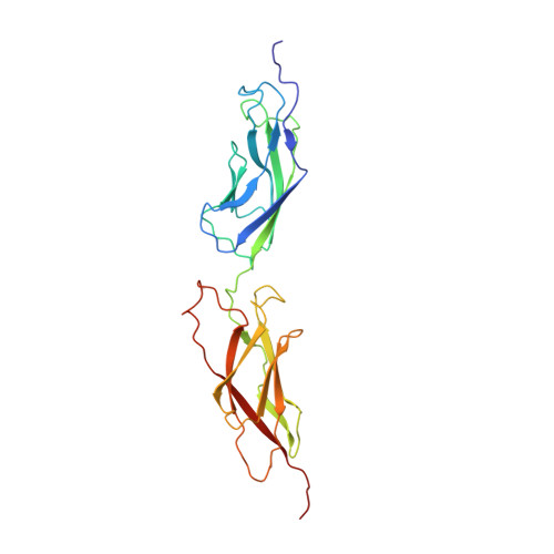
    6SIT

  • PubMed Abstract: 

    The cryo-electron microscopy (cryo-EM) structure of the complex between the trimeric human adenovirus B serotype 3 fibre knob and human desmoglein 2 fragments containing cadherin domains EC2 and EC3 has been published, showing 3:1 and 3:2 complexes. Here, the crystal structure determined at 4.5 Å resolution is presented with one EC2-EC3 desmoglein fragment bound per fibre knob monomer in the asymmetric unit, leading to an apparent 3:3 stoichiometry. However, in concentrated solution the 3:2 complex is predominant, as shown by small-angle X-ray scattering (SAXS), while cryo-EM at lower concentrations showed a majority of the 3:1 complex. Substitution of the calcium ions bound to the desmoglein domains by terbium ions allowed confirmation of the X-ray model using their anomalous scattering and shows that at least one binding site per cluster of calcium ions is intact and exchangeable and, combined with SAXS data, that the cadherin domains are folded even in the distal part that is invisible in the cryo-EM reconstruction.


  • Organizational Affiliation

    Institut de Biologie Structurale (IBS), Université Grenoble Alpes, CNRS, CEA, 71 Avenue des Martyrs, 38000 Grenoble, France.


Macromolecules
Find similar proteins by:  (by identity cutoff)  |  3D Structure
Entity ID: 1
MoleculeChains Sequence LengthOrganismDetailsImage
Fiber protein191Human adenovirus B3Mutation(s): 0 
Gene Names: L5
UniProt
Find proteins for P04501 (Human adenovirus B serotype 3)
Explore P04501 
Go to UniProtKB:  P04501
Entity Groups  
Sequence Clusters30% Identity50% Identity70% Identity90% Identity95% Identity100% Identity
UniProt GroupP04501
Sequence Annotations
Expand
  • Reference Sequence
Find similar proteins by:  (by identity cutoff)  |  3D Structure
Entity ID: 2
MoleculeChains Sequence LengthOrganismDetailsImage
Desmoglein-2B [auth D]238Homo sapiensMutation(s): 0 
Gene Names: DSG2CDHF5
UniProt & NIH Common Fund Data Resources
Find proteins for Q14126 (Homo sapiens)
Explore Q14126 
Go to UniProtKB:  Q14126
PHAROS:  Q14126
GTEx:  ENSG00000046604 
Entity Groups  
Sequence Clusters30% Identity50% Identity70% Identity90% Identity95% Identity100% Identity
UniProt GroupQ14126
Sequence Annotations
Expand
  • Reference Sequence
Experimental Data & Validation

Experimental Data

  • Method: X-RAY DIFFRACTION
  • Resolution: 4.50 Å
  • R-Value Free: 0.370 
  • R-Value Work: 0.367 
  • Space Group: I 21 3
Unit Cell:
Length ( Å )Angle ( ˚ )
a = 146.53α = 90
b = 146.53β = 90
c = 146.53γ = 90
Software Package:
Software NamePurpose
CNSrefinement
PDB_EXTRACTdata extraction
XDSdata reduction
Aimlessdata scaling
MOLREPphasing

Structure Validation

View Full Validation Report


Entry History & Funding Information

Deposition Data


Funding OrganizationLocationGrant Number
French National Research AgencyFranceANR-10-INSB-05-02
French National Research AgencyFranceANR-10-LABX-49-01

Revision History  (Full details and data files)

  • Version 1.0: 2019-12-18
    Type: Initial release
  • Version 1.1: 2024-01-24
    Changes: Data collection, Database references, Derived calculations, Refinement description