6RWL

SIVrcm intasome


Experimental Data Snapshot

  • Method: ELECTRON MICROSCOPY
  • Resolution: 3.36 Å
  • Aggregation State: PARTICLE 
  • Reconstruction Method: SINGLE PARTICLE 

wwPDB Validation   3D Report Full Report


This is version 1.3 of the entry. See complete history


Literature

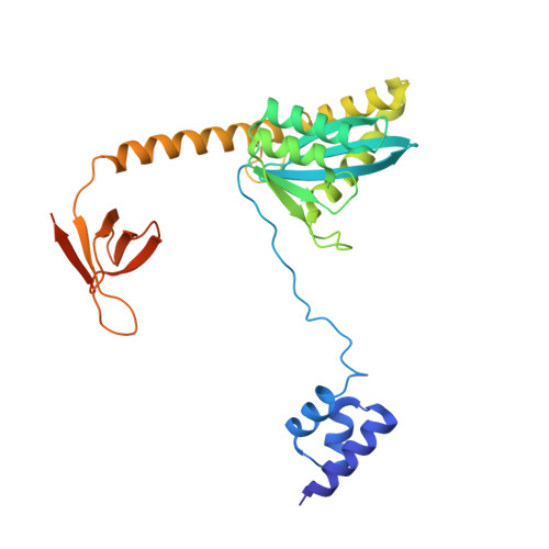
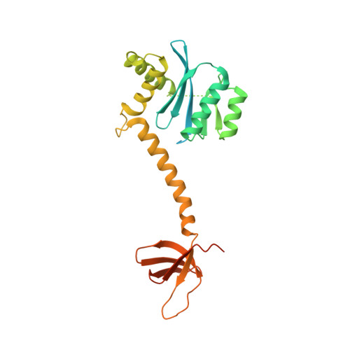
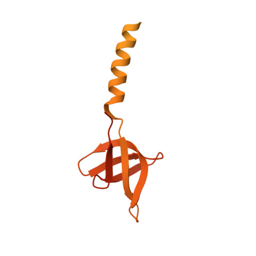
Structural basis of second-generation HIV integrase inhibitor action and viral resistance.

Cook, N.J.Li, W.Berta, D.Badaoui, M.Ballandras-Colas, A.Nans, A.Kotecha, A.Rosta, E.Engelman, A.N.Cherepanov, P.

(2020) Science 367: 806-810

  • DOI: https://doi.org/10.1126/science.aay4919
  • Primary Citation of Related Structures:  
    6RWL, 6RWM, 6RWN, 6RWO

  • PubMed Abstract: 

    Although second-generation HIV integrase strand-transfer inhibitors (INSTIs) are prescribed throughout the world, the mechanistic basis for the superiority of these drugs is poorly understood. We used single-particle cryo-electron microscopy to visualize the mode of action of the advanced INSTIs dolutegravir and bictegravir at near-atomic resolution. Glutamine-148→histidine (Q148H) and glycine-140→serine (G140S) amino acid substitutions in integrase that result in clinical INSTI failure perturb optimal magnesium ion coordination in the enzyme active site. The expanded chemical scaffolds of second-generation compounds mediate interactions with the protein backbone that are critical for antagonizing viruses containing the Q148H and G140S mutations. Our results reveal that binding to magnesium ions underpins a fundamental weakness of the INSTI pharmacophore that is exploited by the virus to engender resistance and provide a structural framework for the development of this class of anti-HIV/AIDS therapeutics.


  • Organizational Affiliation

    Chromatin Structure and Mobile DNA Laboratory, Francis Crick Institute, London NW1 1AT, UK.


Macromolecules

Find similar proteins by:  (by identity cutoff)  |  3D Structure
Entity ID: 1
MoleculeChains Sequence LengthOrganismDetailsImage
Pol protein290Simian immunodeficiency virusMutation(s): 1 
Gene Names: pol
UniProt
Find proteins for E1ANT8 (Simian immunodeficiency virus)
Explore E1ANT8 
Go to UniProtKB:  E1ANT8
Entity Groups  
Sequence Clusters30% Identity50% Identity70% Identity90% Identity95% Identity100% Identity
UniProt GroupE1ANT8
Sequence Annotations
Expand
  • Reference Sequence
Find similar proteins by:  (by identity cutoff)  |  3D Structure
Entity ID: 2
MoleculeChains Sequence LengthOrganismDetailsImage
Pol protein
D, L
289Simian immunodeficiency virusMutation(s): 1 
Gene Names: pol
UniProt
Find proteins for E1ANT8 (Simian immunodeficiency virus)
Explore E1ANT8 
Go to UniProtKB:  E1ANT8
Entity Groups  
Sequence Clusters30% Identity50% Identity70% Identity90% Identity95% Identity100% Identity
UniProt GroupE1ANT8
Sequence Annotations
Expand
  • Reference Sequence
Find similar proteins by:  (by identity cutoff)  |  3D Structure
Entity ID: 3
MoleculeChains Sequence LengthOrganismDetailsImage
Pol proteinE [auth F],
M [auth N]
290Simian immunodeficiency virusMutation(s): 0 
Gene Names: pol
UniProt
Find proteins for E1ANT8 (Simian immunodeficiency virus)
Explore E1ANT8 
Go to UniProtKB:  E1ANT8
Entity Groups  
Sequence Clusters30% Identity50% Identity70% Identity90% Identity95% Identity100% Identity
UniProt GroupE1ANT8
Sequence Annotations
Expand
  • Reference Sequence
Find similar nucleic acids by:  (by identity cutoff)  |  3D Structure
Entity ID: 4
MoleculeChains LengthOrganismImage
DNA (5'-D(P*GP*CP*TP*AP*AP*GP*AP*AP*AP*AP*AP*TP*CP*TP*CP*TP*AP*CP*CP*A)-3')G [auth T],
O [auth W]
30Simian immunodeficiency virus
Sequence Annotations
Expand
  • Reference Sequence
Find similar nucleic acids by:  (by identity cutoff)  |  3D Structure
Entity ID: 5
MoleculeChains LengthOrganismImage
DNA (5'-D(*AP*AP*CP*TP*GP*GP*TP*AP*GP*AP*GP*AP*TP*TP*TP*TP*TP*CP*TP*TP*AP*GP*C)-3')H [auth S],
P [auth Q]
33Simian immunodeficiency virus
Sequence Annotations
Expand
  • Reference Sequence
Experimental Data & Validation

Experimental Data

  • Method: ELECTRON MICROSCOPY
  • Resolution: 3.36 Å
  • Aggregation State: PARTICLE 
  • Reconstruction Method: SINGLE PARTICLE 
EM Software:
TaskSoftware PackageVersion
MODEL REFINEMENTCoot
MODEL REFINEMENTPHENIX1.15.2-3472
RECONSTRUCTIONcryoSPARC2

Structure Validation

View Full Validation Report


Entry History & Funding Information

Deposition Data


Funding OrganizationLocationGrant Number
National Institutes of Health/National Institute of General Medical Sciences (NIH/NIGMS)United StatesGM082251
The Francis Crick InstituteUnited KingdomFC001061

Revision History  (Full details and data files)

  • Version 1.0: 2020-02-05
    Type: Initial release
  • Version 1.1: 2020-02-19
    Changes: Database references
  • Version 1.2: 2020-02-26
    Changes: Database references, Structure summary
  • Version 1.3: 2022-03-30
    Changes: Author supporting evidence, Database references