6R6W

Structure of recombinant human butyrylcholinesterase in complex with a fluorescent NBD-based probe


Experimental Data Snapshot

  • Method: X-RAY DIFFRACTION
  • Resolution: 2.47 Å
  • R-Value Free: 0.227 
  • R-Value Work: 0.182 
  • R-Value Observed: 0.184 

wwPDB Validation   3D Report Full Report


Ligand Structure Quality Assessment 


This is version 2.1 of the entry. See complete history


Literature

Development of potent reversible selective inhibitors of butyrylcholinesterase as fluorescent probes.

Pajk, S.Knez, D.Kosak, U.Zorovic, M.Brazzolotto, X.Coquelle, N.Nachon, F.Colletier, J.P.Zivin, M.Stojan, J.Gobec, S.

(2020) J Enzyme Inhib Med Chem 35: 498-505

  • DOI: https://doi.org/10.1080/14756366.2019.1710502
  • Primary Citation of Related Structures:  
    6R6V, 6R6W, 6RUA

  • PubMed Abstract: 

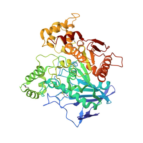
    Brain butyrylcholinesterase (BChE) is an attractive target for drugs designed for the treatment of Alzheimer's disease (AD) in its advanced stages. It also potentially represents a biomarker for progression of this disease. Based on the crystal structure of previously described highly potent, reversible, and selective BChE inhibitors, we have developed the fluorescent probes that are selective towards human BChE. The most promising probes also maintain their inhibition of BChE in the low nanomolar range with high selectivity over acetylcholinesterase. Kinetic studies of probes reveal a reversible mixed inhibition mechanism, with binding of these fluorescent probes to both the free and acylated enzyme. Probes show environment-sensitive emission, and additionally, one of them also shows significant enhancement of fluorescence intensity upon binding to the active site of BChE. Finally, the crystal structures of probes in complex with human BChE are reported, which offer an excellent base for further development of this library of compounds.


  • Organizational Affiliation

    Faculty of Pharmacy, University of Ljubljana, Ljubljana, Slovenia.


Macromolecules
Find similar proteins by:  (by identity cutoff)  |  3D Structure
Entity ID: 1
MoleculeChains Sequence LengthOrganismDetailsImage
Cholinesterase526Homo sapiensMutation(s): 4 
Gene Names: BCHECHE1
EC: 3.1.1.8
UniProt & NIH Common Fund Data Resources
Find proteins for P06276 (Homo sapiens)
Explore P06276 
Go to UniProtKB:  P06276
PHAROS:  P06276
GTEx:  ENSG00000114200 
Entity Groups  
Sequence Clusters30% Identity50% Identity70% Identity90% Identity95% Identity100% Identity
UniProt GroupP06276
Sequence Annotations
Expand
  • Reference Sequence
Oligosaccharides

Help

Entity ID: 2
MoleculeChains Length2D Diagram Glycosylation3D Interactions
alpha-L-fucopyranose-(1-6)-2-acetamido-2-deoxy-beta-D-glucopyranose
B
2N-Glycosylation
Glycosylation Resources
GlyTouCan:  G86851RC
GlyCosmos:  G86851RC
GlyGen:  G86851RC
Entity ID: 3
MoleculeChains Length2D Diagram Glycosylation3D Interactions
2-acetamido-2-deoxy-beta-D-glucopyranose-(1-4)-[alpha-L-fucopyranose-(1-6)]2-acetamido-2-deoxy-beta-D-glucopyranose
C, D
3N-Glycosylation
Glycosylation Resources
GlyTouCan:  G21290RB
GlyCosmos:  G21290RB
GlyGen:  G21290RB
Small Molecules
Ligands 5 Unique
IDChains Name / Formula / InChI Key2D Diagram3D Interactions
JUB (Subject of Investigation/LOI)
Query on JUB

Download Ideal Coordinates CCD File 
H [auth A][7-[4-[2-[naphthalen-2-ylsulfonyl-[[(3~{S})-1-(phenylmethyl)piperidin-1-ium-3-yl]methyl]amino]ethyl]piperazin-4-ium-1-yl]-2,1,3-benzoxadiazol-4-yl]-oxidanyl-oxidanylidene-azanium
C35 H42 N7 O5 S
QBNBLJUWYPBAGF-NDEPHWFRSA-P
NAG
Query on NAG

Download Ideal Coordinates CCD File 
E [auth A],
F [auth A],
G [auth A]
2-acetamido-2-deoxy-beta-D-glucopyranose
C8 H15 N O6
OVRNDRQMDRJTHS-FMDGEEDCSA-N
SO4
Query on SO4

Download Ideal Coordinates CCD File 
J [auth A],
K [auth A],
L [auth A],
M [auth A],
N [auth A]
SULFATE ION
O4 S
QAOWNCQODCNURD-UHFFFAOYSA-L
GOL
Query on GOL

Download Ideal Coordinates CCD File 
I [auth A]GLYCEROL
C3 H8 O3
PEDCQBHIVMGVHV-UHFFFAOYSA-N
DMS
Query on DMS

Download Ideal Coordinates CCD File 
O [auth A]DIMETHYL SULFOXIDE
C2 H6 O S
IAZDPXIOMUYVGZ-UHFFFAOYSA-N
Binding Affinity Annotations 
IDSourceBinding Affinity
JUB Binding MOAD:  6R6W Ki: 18 (nM) from 1 assay(s)
Experimental Data & Validation

Experimental Data

  • Method: X-RAY DIFFRACTION
  • Resolution: 2.47 Å
  • R-Value Free: 0.227 
  • R-Value Work: 0.182 
  • R-Value Observed: 0.184 
  • Space Group: I 4 2 2
Unit Cell:
Length ( Å )Angle ( ˚ )
a = 154.22α = 90
b = 154.22β = 90
c = 127.85γ = 90
Software Package:
Software NamePurpose
PHENIXrefinement
PDB_EXTRACTdata extraction
XDSdata reduction
XDSdata scaling
PHASERphasing

Structure Validation

View Full Validation Report


Ligand Structure Quality Assessment 


Entry History & Funding Information

Deposition Data


Funding OrganizationLocationGrant Number
French Ministry of Armed ForcesFranceNBC-4-C-4210

Revision History  (Full details and data files)

  • Version 1.0: 2020-01-15
    Type: Initial release
  • Version 1.1: 2020-01-22
    Changes: Database references
  • Version 2.0: 2020-07-29
    Type: Remediation
    Reason: Carbohydrate remediation
    Changes: Advisory, Atomic model, Data collection, Derived calculations, Structure summary
  • Version 2.1: 2024-01-24
    Changes: Data collection, Database references, Derived calculations, Refinement description, Structure summary