6PZK

Cryo-EM Structure of the Respiratory Syncytial Virus Polymerase (L) Protein Bound by the Tetrameric Phosphoprotein (P)


Experimental Data Snapshot

  • Method: ELECTRON MICROSCOPY
  • Resolution: 3.20 Å
  • Aggregation State: PARTICLE 
  • Reconstruction Method: SINGLE PARTICLE 

wwPDB Validation   3D Report Full Report


This is version 1.2 of the entry. See complete history


Literature

Structure of the Respiratory Syncytial Virus Polymerase Complex.

Gilman, M.S.A.Liu, C.Fung, A.Behera, I.Jordan, P.Rigaux, P.Ysebaert, N.Tcherniuk, S.Sourimant, J.Eleouet, J.F.Sutto-Ortiz, P.Decroly, E.Roymans, D.Jin, Z.McLellan, J.S.

(2019) Cell 179: 193-204.e14

  • DOI: https://doi.org/10.1016/j.cell.2019.08.014
  • Primary Citation of Related Structures:  
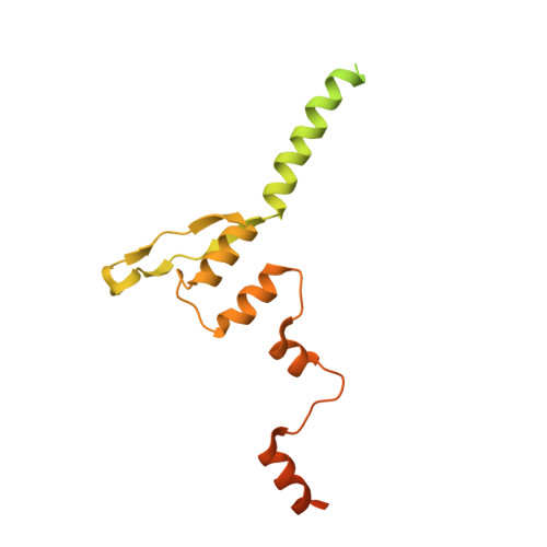
    6PZK

  • PubMed Abstract: 

    Numerous interventions are in clinical development for respiratory syncytial virus (RSV) infection, including small molecules that target viral transcription and replication. These processes are catalyzed by a complex comprising the RNA-dependent RNA polymerase (L) and the tetrameric phosphoprotein (P). RSV P recruits multiple proteins to the polymerase complex and, with the exception of its oligomerization domain, is thought to be intrinsically disordered. Despite their critical roles in RSV transcription and replication, structures of L and P have remained elusive. Here, we describe the 3.2-Å cryo-EM structure of RSV L bound to tetrameric P. The structure reveals a striking tentacular arrangement of P, with each of the four monomers adopting a distinct conformation. The structure also rationalizes inhibitor escape mutants and mutations observed in live-attenuated vaccine candidates. These results provide a framework for determining the molecular underpinnings of RSV replication and transcription and should facilitate the design of effective RSV inhibitors.


  • Organizational Affiliation

    Department of Molecular Biosciences, University of Texas at Austin, Austin, TX 78712, USA.


Macromolecules
Find similar proteins by:  (by identity cutoff)  |  3D Structure
Entity ID: 1
MoleculeChains Sequence LengthOrganismDetailsImage
RNA-directed RNA polymerase L2,201Human respiratory syncytial virus A2Mutation(s): 0 
EC: 2.7.7.48 (PDB Primary Data), 2.1.1.56 (PDB Primary Data), 2.7.7 (PDB Primary Data), 2.7.7.88 (PDB Primary Data)
UniProt
Find proteins for P28887 (Human respiratory syncytial virus A (strain A2))
Explore P28887 
Go to UniProtKB:  P28887
Entity Groups  
Sequence Clusters30% Identity50% Identity70% Identity90% Identity95% Identity100% Identity
UniProt GroupP28887
Sequence Annotations
Expand
  • Reference Sequence
Find similar proteins by:  (by identity cutoff)  |  3D Structure
Entity ID: 2
MoleculeChains Sequence LengthOrganismDetailsImage
Phosphoprotein
B, C, D, E
256Human respiratory syncytial virus A2Mutation(s): 0 
UniProt
Find proteins for P03421 (Human respiratory syncytial virus A (strain A2))
Explore P03421 
Go to UniProtKB:  P03421
Entity Groups  
Sequence Clusters30% Identity50% Identity70% Identity90% Identity95% Identity100% Identity
UniProt GroupP03421
Sequence Annotations
Expand
  • Reference Sequence
Experimental Data & Validation

Experimental Data

  • Method: ELECTRON MICROSCOPY
  • Resolution: 3.20 Å
  • Aggregation State: PARTICLE 
  • Reconstruction Method: SINGLE PARTICLE 
EM Software:
TaskSoftware PackageVersion
RECONSTRUCTIONcryoSPARC

Structure Validation

View Full Validation Report


Entry History 

Deposition Data

Revision History  (Full details and data files)

  • Version 1.0: 2019-09-11
    Type: Initial release
  • Version 1.1: 2019-10-02
    Changes: Data collection, Database references
  • Version 1.2: 2024-03-20
    Changes: Data collection, Database references