6E3R

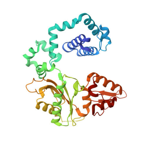
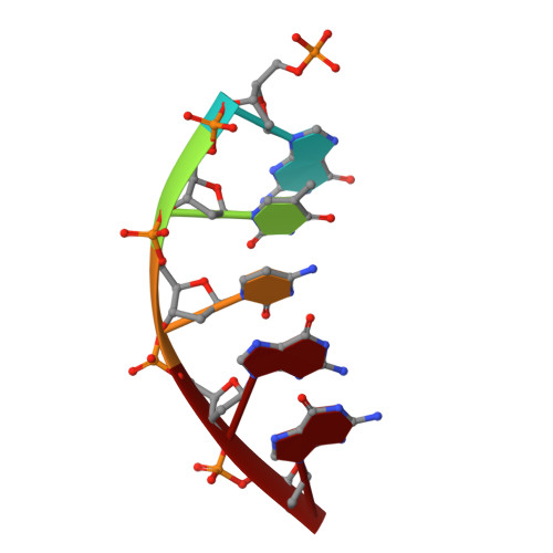
Structure of human DNA polymerase beta complexed with 8OA as the template base in a 1-nucleotide gapped DNA


Experimental Data Snapshot

  • Method: X-RAY DIFFRACTION
  • Resolution: 2.26 Å
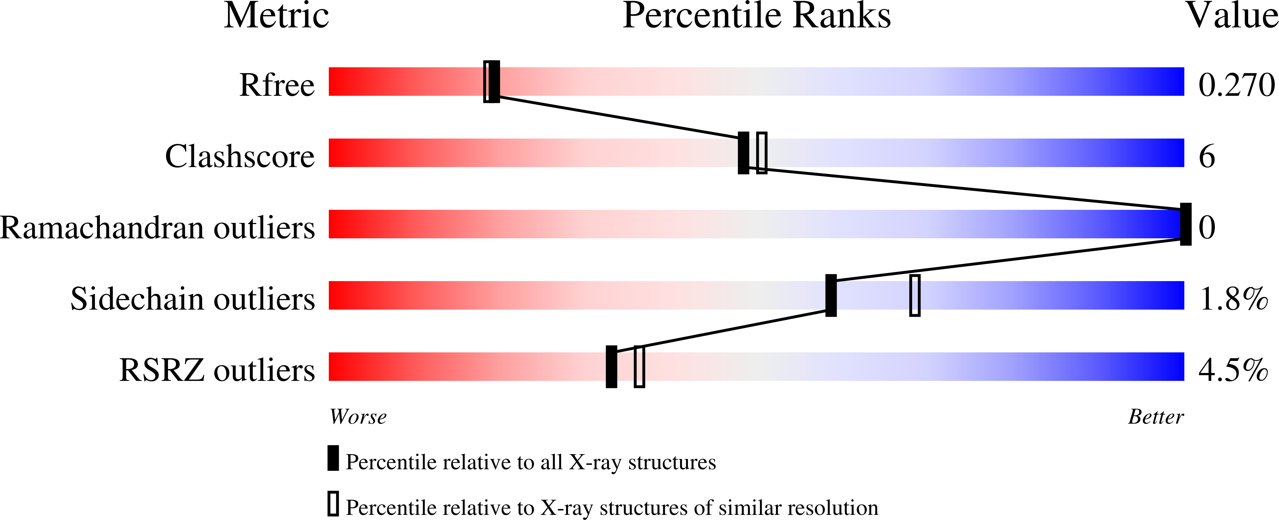
  • R-Value Free: 0.272 
  • R-Value Work: 0.204 
  • R-Value Observed: 0.207 

wwPDB Validation   3D Report Full Report


This is version 1.2 of the entry. See complete history


Literature

Mutagenic Replication of the Major Oxidative Adenine Lesion 7,8-Dihydro-8-oxoadenine by Human DNA Polymerases.

Koag, M.C.Jung, H.Lee, S.

(2019) J Am Chem Soc 141: 4584-4596

  • DOI: https://doi.org/10.1021/jacs.8b08551
  • Primary Citation of Related Structures:  
    6E3R, 6E3V, 6E3W

  • PubMed Abstract: 

    Reactive oxygen species attack DNA to produce 7,8-dihyro-8-oxoguanine (oxoG) and 7,8-dihydro-8-oxoadenine (oxoA) as major lesions. The structural basis for the mutagenicity of oxoG, which induces G to T mutations, is well understood. However, the structural basis for the mutagenic potential of oxoA, which induces A to C mutations, remains poorly understood. To gain insight into oxoA-induced mutagenesis, we conducted kinetic studies of human DNA polymerases β and η replicating across oxoA and structural studies of polβ incorporating dTTP/dGTP opposite oxoA. While polη readily bypassed oxoA, it incorporated dGTP opposite oxoA with a catalytic specificity comparable to that of correct insertion, underscoring the promutagenic nature of the major oxidative adenine lesion. Polη and polβ incorporated dGTP opposite oxoA ∼170-fold and ∼100-fold more efficiently than that opposite dA, respectively, indicating that the 8-oxo moiety greatly facilitated error-prone replication. Crystal structures of polβ showed that, when paired with an incoming dTTP, the templating oxoA adopted an anti conformation and formed Watson-Crick base pair. When paired with dGTP, oxoA adopted a syn conformation and formed a Hoogsteen base pair with Watson-Crick-like geometry, highlighting the dual-coding potential of oxoA. The templating oxoA was stabilized by Lys280-mediated stacking and hydrogen bonds. Overall, these results provide insight into the mutagenic potential and dual-coding nature of the major oxidative adenine lesion.


  • Organizational Affiliation

    The Division of Chemical Biology and Medicinal Chemistry, College of Pharmacy , The University of Texas at Austin , Austin , Texas 78712 , United States.


Macromolecules

Find similar proteins by:  (by identity cutoff)  |  3D Structure
Entity ID: 1
MoleculeChains Sequence LengthOrganismDetailsImage
DNA polymerase beta335Homo sapiensMutation(s): 0 
Gene Names: POLB
EC: 2.7.7.7 (PDB Primary Data), 4.2.99 (PDB Primary Data)
UniProt & NIH Common Fund Data Resources
Find proteins for P06746 (Homo sapiens)
Explore P06746 
Go to UniProtKB:  P06746
PHAROS:  P06746
GTEx:  ENSG00000070501 
Entity Groups  
Sequence Clusters30% Identity50% Identity70% Identity90% Identity95% Identity100% Identity
UniProt GroupP06746
Sequence Annotations
Expand
  • Reference Sequence

Find similar nucleic acids by:  Sequence   |   3D Structure  

Entity ID: 2
MoleculeChains LengthOrganismImage
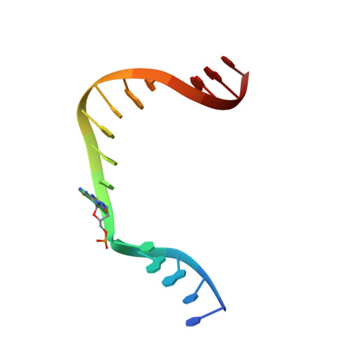
DNA (5'-D(*CP*CP*GP*AP*CP*(A38) P*TP*CP*GP*CP*AP*TP*CP*AP*GP*C)-3')B [auth T]16Homo sapiens
Sequence Annotations
Expand
  • Reference Sequence

Find similar nucleic acids by:  Sequence   |   3D Structure  

Entity ID: 3
MoleculeChains LengthOrganismImage
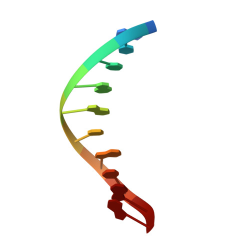
DNA (5'-D(*GP*CP*TP*GP*AP*TP*GP*CP*GP*A)-3')C [auth P]10Homo sapiens
Sequence Annotations
Expand
  • Reference Sequence

Find similar nucleic acids by:  Sequence   |   3D Structure  

Entity ID: 4
MoleculeChains LengthOrganismImage
DNA (5'-D(P*GP*TP*CP*GP*G)-3')5Homo sapiens
Sequence Annotations
Expand
  • Reference Sequence
Experimental Data & Validation

Experimental Data

  • Method: X-RAY DIFFRACTION
  • Resolution: 2.26 Å
  • R-Value Free: 0.272 
  • R-Value Work: 0.204 
  • R-Value Observed: 0.207 
  • Space Group: P 1 21 1
Unit Cell:
Length ( Å )Angle ( ˚ )
a = 54.572α = 90
b = 79.048β = 105.75
c = 54.966γ = 90
Software Package:
Software NamePurpose
DENZOdata reduction
HKL-2000data scaling
MOLREPphasing
PHASERphasing
REFMACrefinement

Structure Validation

View Full Validation Report


Entry History & Funding Information

Deposition Data


Funding OrganizationLocationGrant Number
National Institutes of Health/National Human Genome Research Institute (NIH/NHGRI)United StatesES-26676

Revision History  (Full details and data files)

  • Version 1.0: 2019-04-03
    Type: Initial release
  • Version 1.1: 2019-12-18
    Changes: Author supporting evidence
  • Version 1.2: 2023-10-11
    Changes: Data collection, Database references, Derived calculations, Refinement description