6EO0

Zebrafish Sirt5 in complex with stalled peptidylimidate and bicyclic intermediate of inhibitory compound 29


Experimental Data Snapshot

  • Method: X-RAY DIFFRACTION
  • Resolution: 2.40 Å
  • R-Value Free: 0.305 
  • R-Value Work: 0.253 
  • R-Value Observed: 0.255 

wwPDB Validation   3D Report Full Report


Ligand Structure Quality Assessment 


This is version 1.2 of the entry. See complete history


Literature

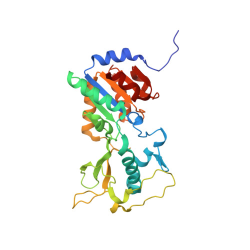
Mechanism-Based Inhibitors of the Human Sirtuin 5 Deacylase: Structure-Activity Relationship, Biostructural, and Kinetic Insight.

Rajabi, N.Auth, M.Troelsen, K.R.Pannek, M.Bhatt, D.P.Fontenas, M.Hirschey, M.D.Steegborn, C.Madsen, A.S.Olsen, C.A.

(2017) Angew Chem Int Ed Engl 56: 14836-14841

  • DOI: https://doi.org/10.1002/anie.201709050
  • Primary Citation of Related Structures:  
    6ENX, 6EO0, 6EQS

  • PubMed Abstract: 

    The sirtuin enzymes are important regulatory deacylases in a variety of biochemical contexts and may therefore be potential therapeutic targets through either activation or inhibition by small molecules. Here, we describe the discovery of the most potent inhibitor of sirtuin 5 (SIRT5) reported to date. We provide rationalization of the mode of binding by solving co-crystal structures of selected inhibitors in complex with both human and zebrafish SIRT5, which provide insight for future optimization of inhibitors with more "drug-like" properties. Importantly, enzyme kinetic evaluation revealed a slow, tight-binding mechanism of inhibition, which is unprecedented for SIRT5. This is important information when applying inhibitors to probe mechanisms in biology.


  • Organizational Affiliation

    Center for Biopharmaceuticals & Department of Drug Design and Pharmacology, University of Copenhagen, Universitetsparken 2, 2100, Copenhagen, Denmark.


Macromolecules
Find similar proteins by:  (by identity cutoff)  |  3D Structure
Entity ID: 1
MoleculeChains Sequence LengthOrganismDetailsImage
NAD-dependent protein deacylase sirtuin-5, mitochondrial
A, B, C, D
277Danio rerioMutation(s): 0 
Gene Names: sirt5si:ch211-121a2.1
EC: 3.5.1
UniProt
Find proteins for Q6DHI5 (Danio rerio)
Explore Q6DHI5 
Go to UniProtKB:  Q6DHI5
Entity Groups  
Sequence Clusters30% Identity50% Identity70% Identity90% Identity95% Identity100% Identity
UniProt GroupQ6DHI5
Sequence Annotations
Expand
  • Reference Sequence
Small Molecules
Ligands 4 Unique
IDChains Name / Formula / InChI Key2D Diagram3D Interactions
BVT
Query on BVT

Download Ideal Coordinates CCD File 
H [auth A],
K [auth B],
N [auth C],
Q [auth D]
3-[[(2~{S},3~{a}~{R},5~{R},6~{R},6~{a}~{R})-5-[[[[(2~{R},3~{S},4~{R},5~{R})-5-(6-aminopurin-9-yl)-3,4-bis(oxidanyl)oxolan-2-yl]methoxy-oxidanyl-phosphoryl]oxy-oxidanyl-phosphoryl]oxymethyl]-2-[[(5~{S})-6-[[(2~{S})-3-(1~{H}-indol-3-yl)-1-oxidanylidene-1-(propan-2-ylamino)propan-2-yl]amino]-6-oxidanylidene-5-(phenylmethoxycarbonylamino)hexyl]amino]-6-oxidanyl-3~{a},5,6,6~{a}-tetrahydrofuro[2,3-d][1,3]oxathiol-2-yl]amino]propanoic acid
C47 H63 N11 O19 P2 S
YXTVLLWQDXVSFE-WDVUSYHVSA-N
BV8
Query on BV8

Download Ideal Coordinates CCD File 
G [auth A]3-[[(~{Z})-~{C}-[(2~{R},3~{R},4~{S},5~{R})-5-[[[[(2~{R},3~{S},4~{R},5~{R})-5-(6-aminopurin-9-yl)-3,4-bis(oxidanyl)oxolan-2-yl]methoxy-oxidanyl-phosphoryl]oxy-oxidanyl-phosphoryl]oxymethyl]-3,4-bis(oxidanyl)oxolan-2-yl]sulfanyl-~{N}-[(5~{S})-6-[[(2~{S})-3-(1~{H}-indol-3-yl)-1-oxidanylidene-1-(propan-2-ylamino)propan-2-yl]amino]-6-oxidanylidene-5-(phenylmethoxycarbonylamino)hexyl]carbonimidoyl]amino]propanoic acid
C47 H63 N11 O19 P2 S
GGZPCSBRGOFORP-PVLZSEPSSA-N
ZN
Query on ZN

Download Ideal Coordinates CCD File 
E [auth A],
I [auth B],
L [auth C],
O [auth D]
ZINC ION
Zn
PTFCDOFLOPIGGS-UHFFFAOYSA-N
EDO
Query on EDO

Download Ideal Coordinates CCD File 
F [auth A],
J [auth B],
M [auth C],
P [auth D]
1,2-ETHANEDIOL
C2 H6 O2
LYCAIKOWRPUZTN-UHFFFAOYSA-N
Experimental Data & Validation

Experimental Data

  • Method: X-RAY DIFFRACTION
  • Resolution: 2.40 Å
  • R-Value Free: 0.305 
  • R-Value Work: 0.253 
  • R-Value Observed: 0.255 
  • Space Group: P 1 21 1
Unit Cell:
Length ( Å )Angle ( ˚ )
a = 64.93α = 90
b = 113.59β = 103.24
c = 72.31γ = 90
Software Package:
Software NamePurpose
REFMACrefinement
XDSdata reduction
XDSdata scaling
PHASERphasing

Structure Validation

View Full Validation Report


Ligand Structure Quality Assessment 


Entry History & Funding Information

Deposition Data


Funding OrganizationLocationGrant Number
OberfrankenstiftungGermany04115

Revision History  (Full details and data files)

  • Version 1.0: 2017-11-01
    Type: Initial release
  • Version 1.1: 2017-11-22
    Changes: Database references
  • Version 1.2: 2024-01-17
    Changes: Data collection, Database references, Refinement description