6BT4

Crystal structure of the SLH domain of Sap from Bacillus anthracis in complex with a pyruvylated SCWP unit


Experimental Data Snapshot

  • Method: X-RAY DIFFRACTION
  • Resolution: 2.31 Å
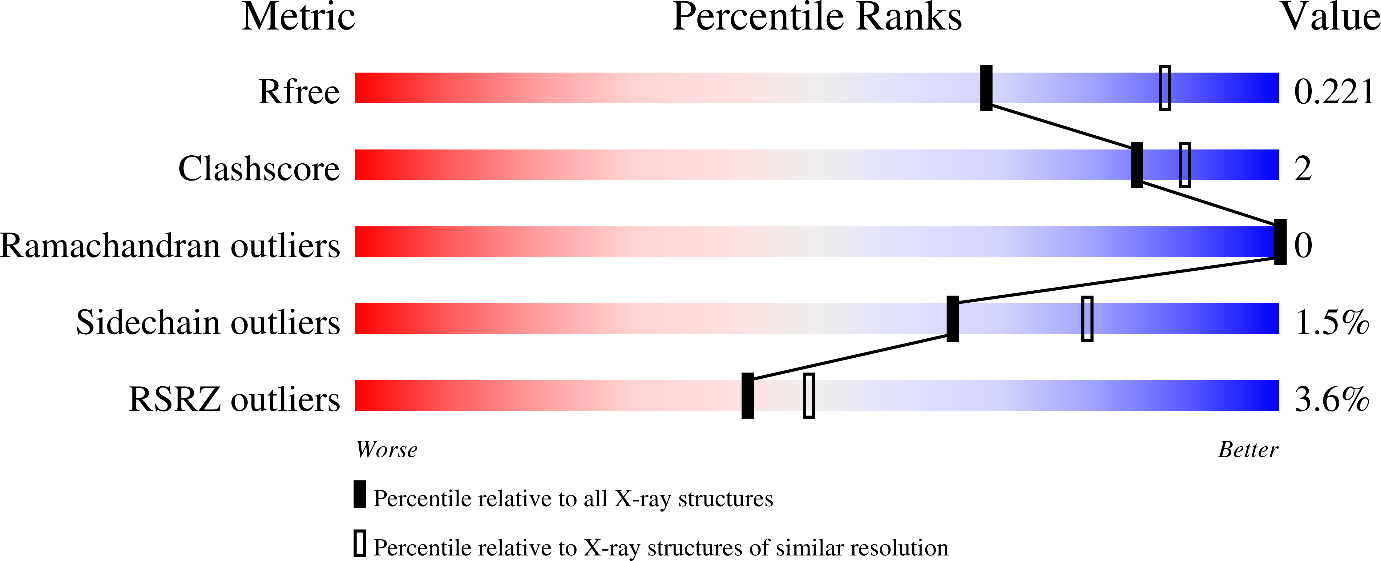
  • R-Value Free: 0.218 
  • R-Value Work: 0.185 
  • R-Value Observed: 0.188 

wwPDB Validation   3D Report Full Report


Ligand Structure Quality Assessment 


This is version 1.3 of the entry. See complete history


Literature

Molecular Basis for the Attachment of S-Layer Proteins to the Cell Wall of Bacillus anthracis.

Sychantha, D.Chapman, R.N.Bamford, N.C.Boons, G.J.Howell, P.L.Clarke, A.J.

(2018) Biochemistry 57: 1949-1953

  • DOI: https://doi.org/10.1021/acs.biochem.8b00060
  • Primary Citation of Related Structures:  
    6BT4

  • PubMed Abstract: 

    Bacterial surface (S) layers are paracrystalline arrays of protein assembled on the bacterial cell wall that serve as protective barriers and scaffolds for housekeeping enzymes and virulence factors. The attachment of S-layer proteins to the cell walls of the Bacillus cereus sensu lato, which includes the pathogen Bacillus anthracis, occurs through noncovalent interactions between their S-layer homology domains and secondary cell wall polysaccharides. To promote these interactions, it is presumed that the terminal N-acetylmannosamine (ManNAc) residues of the secondary cell wall polysaccharides must be ketal-pyruvylated. For a few specific S-layer proteins, the O-acetylation of the penultimate N-acetylglucosamine (GlcNAc) is also required. Herein, we present the X-ray crystal structure of the SLH domain of the major surface array protein Sap from B. anthracis in complex with 4,6- O-ketal-pyruvyl-β-ManNAc-(1,4)-β-GlcNAc-(1,6)-α-GlcN. This structure reveals for the first time that the conserved terminal SCWP unit is the direct ligand for the SLH domain. Furthermore, we identify key binding interactions that account for the requirement of 4,6- O-ketal-pyruvyl-ManNAc while revealing the insignificance of the O-acetylation on the GlcNAc residue for recognition by Sap.


  • Organizational Affiliation

    Department of Molecular and Cellular Biology , University of Guelph , Guelph , ON N1G 2X1 , Canada.


Macromolecules
Find similar proteins by:  (by identity cutoff)  |  3D Structure
Entity ID: 1
MoleculeChains Sequence LengthOrganismDetailsImage
S-layer protein sap202Bacillus anthracisMutation(s): 0 
Gene Names: sapBA_0885GBAA_0885BAS0841
UniProt
Find proteins for P49051 (Bacillus anthracis)
Explore P49051 
Go to UniProtKB:  P49051
Entity Groups  
Sequence Clusters30% Identity50% Identity70% Identity90% Identity95% Identity100% Identity
UniProt GroupP49051
Sequence Annotations
Expand
  • Reference Sequence
Small Molecules
Ligands 2 Unique
IDChains Name / Formula / InChI Key2D Diagram3D Interactions
KPM (Subject of Investigation/LOI)
Query on KPM

Download Ideal Coordinates CCD File 
B [auth A]2-(acetylamino)-4-O-{2-(acetylamino)-4,6-O-[(1S)-1-carboxyethylidene]-2-deoxy-beta-D-mannopyranosyl}-2-deoxy-beta-D-glucopyranose
C19 H30 N2 O13
ZDEWQEOMIIQXJL-JRZDJGMASA-N
SO4
Query on SO4

Download Ideal Coordinates CCD File 
C [auth A],
D [auth A]
SULFATE ION
O4 S
QAOWNCQODCNURD-UHFFFAOYSA-L
Experimental Data & Validation

Experimental Data

  • Method: X-RAY DIFFRACTION
  • Resolution: 2.31 Å
  • R-Value Free: 0.218 
  • R-Value Work: 0.185 
  • R-Value Observed: 0.188 
  • Space Group: P 41 21 2
Unit Cell:
Length ( Å )Angle ( ˚ )
a = 76.56α = 90
b = 76.56β = 90
c = 97.379γ = 90
Software Package:
Software NamePurpose
PHENIXrefinement
XDSdata scaling
Cootmodel building
PHENIXphasing
XDSdata processing
XDSdata reduction

Structure Validation

View Full Validation Report


Ligand Structure Quality Assessment 


Entry History & Funding Information

Deposition Data


Funding OrganizationLocationGrant Number
Canadian Institutes of Health Research (CIHR)CanadaTGC 114045
Canadian Institutes of Health Research (CIHR)CanadaMOP 43998

Revision History  (Full details and data files)

  • Version 1.0: 2018-03-21
    Type: Initial release
  • Version 1.1: 2018-04-18
    Changes: Data collection, Database references
  • Version 1.2: 2020-01-08
    Changes: Author supporting evidence
  • Version 1.3: 2023-10-04
    Changes: Data collection, Database references, Refinement description