Catalytic Role of Conserved Asparagine, Glutamine, Serine, and Tyrosine Residues in Isoprenoid Biosynthesis Enzymes.
Malwal, S.R., Gao, J., Hu, X., Yang, Y., Liu, W., Huang, J.W., Ko, T.P., Li, L., Chen, C.C., O'Dowd, B., Khade, R.L., Zhang, Y., Zhang, Y., Oldfield, E., Guo, R.T.(2018) ACS Catal 8: 4299-4312
- PubMed: 30345154
- DOI: https://doi.org/10.1021/acscatal.8b00543
- Primary Citation of Related Structures:
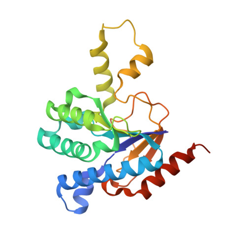
5YGJ, 5YGK - PubMed Abstract:
We report the results of an investigation into the catalytic role of highly conserved amide (asparagine, glutamine) and OH-containing (serine, tyrosine) residues in several prenyltransferases. We first obtained the X-ray structure of cyclolavandulyl diphosphate synthase containing two molecules of the substrate analog dimethylallyl ( S )-thiolodiphosphate (DMASPP). The two molecules have similar diphosphate group orientations to those seen in other ζ-fold ( cis - head-to-tail and head-to-middle) prenyltransferases with one diphosphate moiety forming a bidentate chelate with Mg 2+ in the so-called S1 site (which is typically the allylic binding site in ζ-fold proteins) while the second diphosphate binds to Mg 2+ in the so-called S2 site (which is typically the homoallylic binding site in ζ-fold proteins) via a single P1O1 oxygen. The latter interaction can facilitate direct phosphate-mediated proton abstraction via P1O2, or more likely by an indirect mechanism in which P1O2 stabilizes a basic asparagine species that removes H + , which is then eliminated via an Asn-Ser shuttle. The universal occurrence of Asn-Ser pairs in ζ-fold proteins leads to the idea that the highly conserved amide (Asn, Gln) and OH-containing (Tyr) residues seen in many "head-to-head" prenyltransferases such as squalene and dehydrosqualene synthase might play similar roles, in H + elimination. Structural, bioinformatics and mutagenesis investigations indeed indicate an important role of these residues in catalysis, with the results of density functional theory calculations showing that Asn bound to Mg 2+ can act as a general (imine-like) base, while Gln, Tyr and H 2 O form a proton channel that is adjacent to the conventional (Asp-rich) "active site". Taken together, our results lead to mechanisms of proton-elimination from carbocations in numerous prenyltransferases in which neutral species (Asn, Gln, Ser, Tyr, H 2 O) act as proton shuttles, complementing the more familiar roles of acidic groups (in Asp and Glu) that bind to Mg 2+ , and basic groups (primarily Arg) that bind to diphosphates, in isoprenoid biosynthesis.
Organizational Affiliation:
Department of Chemistry, University of Illinois, Urbana, IL 61801, USA.