5TL9

crystal structure of mPGES-1 bound to inhibitor


Experimental Data Snapshot

  • Method: X-RAY DIFFRACTION
  • Resolution: 1.20 Å
  • R-Value Free: 0.168 
  • R-Value Work: 0.152 
  • R-Value Observed: 0.153 

wwPDB Validation   3D Report Full Report


Ligand Structure Quality Assessment 


This is version 1.4 of the entry. See complete history


Literature

Discovery and characterization of [(cyclopentyl)ethyl]benzoic acid inhibitors of microsomal prostaglandin E synthase-1.

Partridge, K.M.Antonysamy, S.Bhattachar, S.N.Chandrasekhar, S.Fisher, M.J.Fretland, A.Gooding, K.Harvey, A.Hughes, N.E.Kuklish, S.L.Luz, J.G.Manninen, P.R.McGee, J.E.Mudra, D.R.Navarro, A.Norman, B.H.Quimby, S.J.Schiffler, M.A.Sloan, A.V.Warshawsky, A.M.Weller, J.M.York, J.S.Yu, X.P.

(2017) Bioorg Med Chem Lett 27: 1478-1483

  • DOI: https://doi.org/10.1016/j.bmcl.2016.11.011
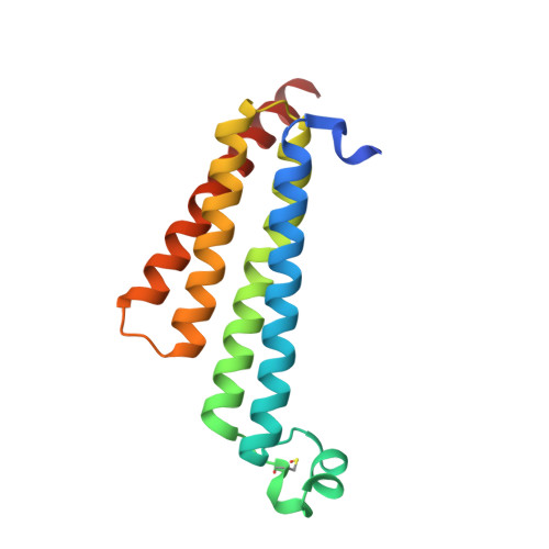
  • Primary Citation of Related Structures:  
    5T36, 5T37, 5TL9

  • PubMed Abstract: 

    We describe a novel class of acidic mPGES-1 inhibitors with nanomolar enzymatic and human whole blood (HWB) potency. Rational design in conjunction with structure-based design led initially to the identification of anthranilic acid 5, an mPGES-1 inhibitor with micromolar HWB potency. Structural modifications of 5 improved HWB potency by over 1000×, reduced CYP2C9 single point inhibition, and improved rat clearance, which led to the selection of [(cyclopentyl)ethyl]benzoic acid compound 16 for clinical studies. Compound 16 showed an IC 80 of 24nM for inhibition of PGE 2 formation in vitro in LPS-stimulated HWB. A single oral dose resulted in plasma concentrations of 16 that exceeded its HWB IC 80 in both rat (5mg/kg) and dog (3mg/kg) for over twelve hours.


  • Organizational Affiliation

    Lilly Research Laboratories, A Division of Eli Lilly and Company, Indianapolis, IN 46285, USA. Electronic address: Partridge_Katherine@lilly.com.


Macromolecules
Find similar proteins by:  (by identity cutoff)  |  3D Structure
Entity ID: 1
MoleculeChains Sequence LengthOrganismDetailsImage
Prostaglandin E synthase154Homo sapiensMutation(s): 0 
Gene Names: PTGESMGST1L1MPGES1PGESPIG12
EC: 5.3.99.3
Membrane Entity: Yes 
UniProt & NIH Common Fund Data Resources
Find proteins for O14684 (Homo sapiens)
Explore O14684 
Go to UniProtKB:  O14684
PHAROS:  O14684
GTEx:  ENSG00000148344 
Entity Groups  
Sequence Clusters30% Identity50% Identity70% Identity90% Identity95% Identity100% Identity
UniProt GroupO14684
Sequence Annotations
Expand
  • Reference Sequence
Small Molecules
Ligands 6 Unique
IDChains Name / Formula / InChI Key2D Diagram3D Interactions
7DN
Query on 7DN

Download Ideal Coordinates CCD File 
B [auth A]2-{2-[(1S,2S)-2-{[1-(8-methylquinolin-2-yl)piperidine-4-carbonyl]amino}cyclopentyl]ethyl}benzoic acid
C30 H35 N3 O3
NLKZRAQTODORFW-NVQXNPDNSA-N
GSH
Query on GSH

Download Ideal Coordinates CCD File 
C [auth A]GLUTATHIONE
C10 H17 N3 O6 S
RWSXRVCMGQZWBV-WDSKDSINSA-N
BOG
Query on BOG

Download Ideal Coordinates CCD File 
E [auth A]octyl beta-D-glucopyranoside
C14 H28 O6
HEGSGKPQLMEBJL-RKQHYHRCSA-N
JZR
Query on JZR

Download Ideal Coordinates CCD File 
D [auth A]hexyl beta-D-glucopyranoside
C12 H24 O6
JVAZJLFFSJARQM-RMPHRYRLSA-N
PG4
Query on PG4

Download Ideal Coordinates CCD File 
F [auth A],
G [auth A]
TETRAETHYLENE GLYCOL
C8 H18 O5
UWHCKJMYHZGTIT-UHFFFAOYSA-N
PEG
Query on PEG

Download Ideal Coordinates CCD File 
H [auth A]DI(HYDROXYETHYL)ETHER
C4 H10 O3
MTHSVFCYNBDYFN-UHFFFAOYSA-N
Modified Residues  1 Unique
IDChains TypeFormula2D DiagramParent
CSO
Query on CSO
A
L-PEPTIDE LINKINGC3 H7 N O3 SCYS
Binding Affinity Annotations 
IDSourceBinding Affinity
7DN BindingDB:  5TL9 IC50: min: 1, max: 6 (nM) from 2 assay(s)
Binding MOAD:  5TL9 IC50: 1 (nM) from 1 assay(s)
Experimental Data & Validation

Experimental Data

  • Method: X-RAY DIFFRACTION
  • Resolution: 1.20 Å
  • R-Value Free: 0.168 
  • R-Value Work: 0.152 
  • R-Value Observed: 0.153 
  • Space Group: H 3
Unit Cell:
Length ( Å )Angle ( ˚ )
a = 77.318α = 90
b = 77.318β = 90
c = 122.954γ = 120
Software Package:
Software NamePurpose
REFMACrefinement
iMOSFLMdata reduction
SCALAdata scaling
PHASERphasing

Structure Validation

View Full Validation Report


Ligand Structure Quality Assessment 


Entry History & Funding Information

Deposition Data


Funding OrganizationLocationGrant Number
Department of Energy (DOE, United States)United StatesDE-AC02-06CH11357

Revision History  (Full details and data files)

  • Version 1.0: 2017-03-01
    Type: Initial release
  • Version 1.1: 2017-03-15
    Changes: Database references
  • Version 1.2: 2017-09-06
    Changes: Author supporting evidence
  • Version 1.3: 2019-12-04
    Changes: Author supporting evidence
  • Version 1.4: 2020-07-29
    Type: Remediation
    Reason: Carbohydrate remediation
    Changes: Data collection, Derived calculations, Structure summary