5CAD

Crystal structure of the vicilin from Solanum melongena revealed existence of different anionic ligands in structurally similar pockets


Experimental Data Snapshot

  • Method: X-RAY DIFFRACTION
  • Resolution: 1.49 Å
  • R-Value Free: 0.210 
  • R-Value Work: 0.199 

wwPDB Validation   3D Report Full Report


This is version 1.1 of the entry. See complete history

Re-refinement Note

A newer entry is available that reflects an alternative modeling of the original data: 5VF5


Literature

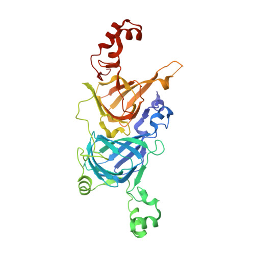
Crystal structure of the vicilin from Solanum melongena reveals existence of different anionic ligands in structurally similar pockets

Jain, A.Kumar, A.Salunke, D.M.

(2016) Sci Rep 6: 23600-23600

  • DOI: https://doi.org/10.1038/srep23600
  • Primary Citation of Related Structures:  
    5CAD

  • PubMed Abstract: 

    Crystal structure of a vicilin, SM80.1, was determined towards exploring its possible physiological functions. The protein was purified from Solanum melongena by combination of ammonium sulphate fractionation and size exclusion chromatography. Structure was determined ab initio at resolution of 1.5 Å by X-ray crystallography showing the three-dimensional topology of the trimeric protein. Each monomer of SM80.1 consists of two similar domains with hydrophobic binding pocket and each accommodating different ligands, i.e. acetate and pyroglutamate. The relatively high stability of these independent anionic ligands in similar pockets indicated a strict requirement of stabilization by hydrogen bonds with the charged residues, suggesting a degree of plasticity within the binding pocket. Comparison of SM80.1 structure with those of other 7S vicilins indicated conservation of putative binding pocket for anionic ligands. Here we propose the possibility of trapping of these ligands in the protein for their requirement in the metabolic processes.


  • Organizational Affiliation

    Regional Centre for Biotechnology, Faridabad-121001, India.


Macromolecules
Find similar proteins by:  (by identity cutoff)  |  3D Structure
Entity ID: 1
MoleculeChains Sequence LengthOrganismDetailsImage
SM80.1 Vicilin392Solanum melongenaMutation(s): 0 
UniProt
Find proteins for A0A158RFR1 (Solanum melongena)
Explore A0A158RFR1 
Go to UniProtKB:  A0A158RFR1
Entity Groups  
Sequence Clusters30% Identity50% Identity70% Identity90% Identity95% Identity100% Identity
UniProt GroupA0A158RFR1
Sequence Annotations
Expand
  • Reference Sequence
Experimental Data & Validation

Experimental Data

  • Method: X-RAY DIFFRACTION
  • Resolution: 1.49 Å
  • R-Value Free: 0.210 
  • R-Value Work: 0.199 
  • Space Group: H 3 2
Unit Cell:
Length ( Å )Angle ( ˚ )
a = 119.33α = 90
b = 119.33β = 90
c = 158.19γ = 120
Software Package:
Software NamePurpose
CNSrefinement
PDB_EXTRACTdata extraction
PHENIXphasing
Cootmodel building
iMOSFLMdata processing
MxCuBEdata collection

Structure Validation

View Full Validation Report


Entry History 

Deposition Data

Revision History  (Full details and data files)

  • Version 1.0: 2016-04-06
    Type: Initial release
  • Version 1.1: 2024-03-20
    Changes: Data collection, Database references, Derived calculations