5A0F

Crystal structure of Yersinia Afp18-modified RhoA


Experimental Data Snapshot

  • Method: X-RAY DIFFRACTION
  • Resolution: 2.00 Å
  • R-Value Free: 0.213 
  • R-Value Work: 0.205 
  • R-Value Observed: 0.206 

wwPDB Validation   3D Report Full Report


Ligand Structure Quality Assessment 


This is version 2.1 of the entry. See complete history


Literature

Tyrosine glycosylation of Rho by Yersinia toxin impairs blastomere cell behaviour in zebrafish embryos.

Jank, T.Eckerle, S.Steinemann, M.Trillhaase, C.Schimpl, M.Wiese, S.van Aalten, D.M.Driever, W.Aktories, K.

(2015) Nat Commun 6: 7807-7807

  • DOI: https://doi.org/10.1038/ncomms8807
  • Primary Citation of Related Structures:  
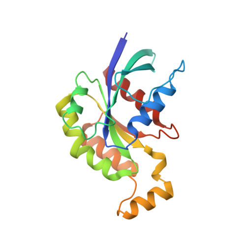
    5A0F

  • PubMed Abstract: 

    Yersinia species cause zoonotic infections, including enterocolitis and plague. Here we studied Yersinia ruckeri antifeeding prophage 18 (Afp18), the toxin component of the phage tail-derived protein translocation system Afp, which causes enteric redmouth disease in salmonid fish species. Here we show that microinjection of the glycosyltransferase domain Afp18(G) into zebrafish embryos blocks cytokinesis, actin-dependent motility and cell blebbing, eventually abrogating gastrulation. In zebrafish ZF4 cells, Afp18(G) depolymerizes actin stress fibres by mono-O-GlcNAcylation of RhoA at tyrosine-34; thereby Afp18(G) inhibits RhoA activation by guanine nucleotide exchange factors, and blocks RhoA, but not Rac and Cdc42 downstream signalling. The crystal structure of tyrosine-GlcNAcylated RhoA reveals an open conformation of the effector loop distinct from recently described structures of GDP- or GTP-bound RhoA. Unravelling of the molecular mechanism of the toxin component Afp18 as glycosyltransferase opens new perspectives in studies of phage tail-derived protein translocation systems, which are preserved from archaea to human pathogenic prokaryotes.


  • Organizational Affiliation

    Institute of Experimental and Clinical Pharmacology and Toxicology, Albert-Ludwigs-University Freiburg, D-79104 Freiburg, Germany.


Macromolecules
Find similar proteins by:  (by identity cutoff)  |  3D Structure
Entity ID: 1
MoleculeChains Sequence LengthOrganismDetailsImage
TRANSFORMING PROTEIN RHOA181Homo sapiensMutation(s): 0 
UniProt & NIH Common Fund Data Resources
Find proteins for P61586 (Homo sapiens)
Explore P61586 
Go to UniProtKB:  P61586
PHAROS:  P61586
GTEx:  ENSG00000067560 
Entity Groups  
Sequence Clusters30% Identity50% Identity70% Identity90% Identity95% Identity100% Identity
UniProt GroupP61586
Sequence Annotations
Expand
  • Reference Sequence
Small Molecules
Experimental Data & Validation

Experimental Data

  • Method: X-RAY DIFFRACTION
  • Resolution: 2.00 Å
  • R-Value Free: 0.213 
  • R-Value Work: 0.205 
  • R-Value Observed: 0.206 
  • Space Group: P 43 21 2
Unit Cell:
Length ( Å )Angle ( ˚ )
a = 91.392α = 90
b = 91.392β = 90
c = 56.628γ = 90
Software Package:
Software NamePurpose
REFMACrefinement
HKL-3000data reduction
SCALEPACKdata scaling
MOLREPphasing

Structure Validation

View Full Validation Report


Ligand Structure Quality Assessment 


Entry History 

Deposition Data

Revision History  (Full details and data files)

  • Version 1.0: 2015-07-22
    Type: Initial release
  • Version 1.1: 2015-07-29
    Changes: Database references
  • Version 1.2: 2017-06-28
    Changes: Data collection
  • Version 1.3: 2017-12-13
    Changes: Database references, Source and taxonomy, Structure summary
  • Version 1.4: 2019-03-13
    Changes: Data collection, Database references, Derived calculations, Experimental preparation
  • Version 2.0: 2020-07-29
    Type: Remediation
    Reason: Carbohydrate remediation
    Changes: Atomic model, Data collection, Derived calculations, Other, Structure summary
  • Version 2.1: 2024-01-10
    Changes: Data collection, Database references, Refinement description, Structure summary