5Y37

Crystal structure of GBS GAPDH


Experimental Data Snapshot

  • Method: X-RAY DIFFRACTION
  • Resolution: 1.36 Å
  • R-Value Free: 0.185 
  • R-Value Work: 0.166 
  • R-Value Observed: 0.167 

wwPDB Validation   3D Report Full Report


Ligand Structure Quality Assessment 


This is version 1.2 of the entry. See complete history


Literature

High-resolution crystal structure of Streptococcus agalactiae glyceraldehyde-3-phosphate dehydrogenase.

Zhou, K.Fan, X.Li, Y.Zhang, C.Jin, T.

(2018) Acta Crystallogr F Struct Biol Commun 74: 236-244

  • DOI: https://doi.org/10.1107/S2053230X18003801
  • Primary Citation of Related Structures:  
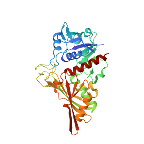
    5Y37

  • PubMed Abstract: 

    Glyceraldehyde-3-phosphate dehydrogenase (GAPDH) is a multifunctional enzyme that plays critical roles in bacterial pathogenesis in some pathogenic bacteria. In this study, the crystal structure of group B streptococcus GAPDH was determined at 1.36 Å resolution. The structure contained an asymmetric mixed holo tetramer, with two NAD ligands bound to two protomers. Further structural analysis identified interesting phosphate ion-binding sites, which shed light on its catalytic mechanism.


  • Organizational Affiliation

    Laboratory of Structural Immunology, CAS Key Laboratory of Innate Immunity and Chronic Disease, CAS Center for Excellence in Molecular Cell Science, School of Life Sciences and Medical Center, University of Science and Technology of China, Hefei, Anhui 230027, People's Republic of China.


Macromolecules
Find similar proteins by:  (by identity cutoff)  |  3D Structure
Entity ID: 1
MoleculeChains Sequence LengthOrganismDetailsImage
Glyceraldehyde-3-phosphate dehydrogenase
A, B, C, D
344Streptococcus agalactiae NEM316Mutation(s): 0 
Gene Names: gbs1811
EC: 1.2.1
UniProt
Find proteins for Q8E3E8 (Streptococcus agalactiae serotype III (strain NEM316))
Explore Q8E3E8 
Go to UniProtKB:  Q8E3E8
Entity Groups  
Sequence Clusters30% Identity50% Identity70% Identity90% Identity95% Identity100% Identity
UniProt GroupQ8E3E8
Sequence Annotations
Expand
  • Reference Sequence
Small Molecules
Ligands 2 Unique
IDChains Name / Formula / InChI Key2D Diagram3D Interactions
NAD
Query on NAD

Download Ideal Coordinates CCD File 
E [auth A],
G [auth B]
NICOTINAMIDE-ADENINE-DINUCLEOTIDE
C21 H27 N7 O14 P2
BAWFJGJZGIEFAR-NNYOXOHSSA-N
SO4
Query on SO4

Download Ideal Coordinates CCD File 
F [auth A]
H [auth B]
I [auth C]
J [auth C]
K [auth C]
F [auth A],
H [auth B],
I [auth C],
J [auth C],
K [auth C],
L [auth C],
M [auth C],
N [auth D],
O [auth D],
P [auth D],
Q [auth D]
SULFATE ION
O4 S
QAOWNCQODCNURD-UHFFFAOYSA-L
Experimental Data & Validation

Experimental Data

  • Method: X-RAY DIFFRACTION
  • Resolution: 1.36 Å
  • R-Value Free: 0.185 
  • R-Value Work: 0.166 
  • R-Value Observed: 0.167 
  • Space Group: P 21 21 2
Unit Cell:
Length ( Å )Angle ( ˚ )
a = 144.3α = 90
b = 156.74β = 90
c = 67.82γ = 90
Software Package:
Software NamePurpose
PHENIXrefinement
XDSdata reduction
XDSdata scaling
PHASERphasing

Structure Validation

View Full Validation Report


Ligand Structure Quality Assessment 


Entry History 

Deposition Data

Revision History  (Full details and data files)

  • Version 1.0: 2018-04-18
    Type: Initial release
  • Version 1.1: 2020-01-01
    Changes: Database references
  • Version 1.2: 2023-11-22
    Changes: Data collection, Database references, Refinement description