4MGM

Crystal structure of the in vitro transcribed G. kaustophilus tRNA-Gly

  • Classification: RNA
  • Mutation(s): No 

  • Deposited: 2013-08-28 Released: 2013-10-23 
  • Deposition Author(s): Grigg, J.C., Ke, A.

Experimental Data Snapshot

  • Method: X-RAY DIFFRACTION
  • Resolution: 3.20 Å
  • R-Value Free: 0.261 
  • R-Value Work: 0.248 
  • R-Value Observed: 0.248 

wwPDB Validation   3D Report Full Report


This is version 1.3 of the entry. See complete history


Literature

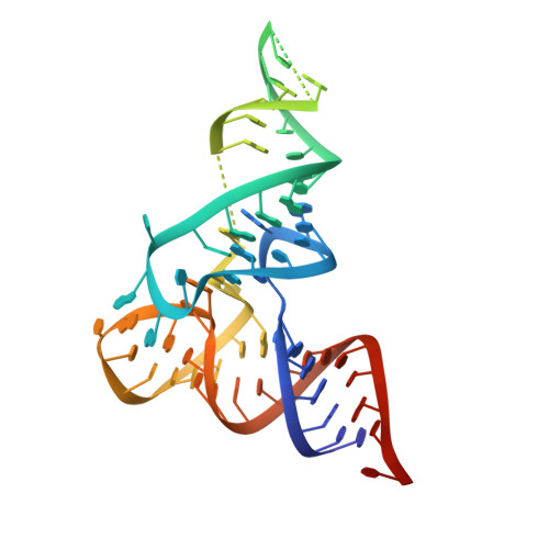
Structural Determinants for Geometry and Information Decoding of tRNA by T Box Leader RNA.

Grigg, J.C.Ke, A.

(2013) Structure 21: 2025-2032

  • DOI: https://doi.org/10.1016/j.str.2013.09.001
  • Primary Citation of Related Structures:  
    4MGM, 4MGN

  • PubMed Abstract: 

    T box riboswitches are cis-acting RNA elements that bind to tRNA and sense its aminoacylation state to influence gene expression. Here, we present the 3.2 Å resolution X-ray crystal structures of the T box Stem I-tRNA complex and tRNA, in isolation. T box Stem I forms an arched conformation with extensive intermolecular contacts to two key points of tRNA, the anticodon and D/T-loops. Free and complexed tRNA exist in significantly different conformations, with the contacts stabilizing flexible D/T-loops and a rearrangement of the D-loop. Using a designed T box RNA/tRNA system, we demonstrate that the T box riboswitch monitors the length and orientation of two essential contacts. Length or orientation mismatches engineered into the T box riboswitch and tRNA disrupt the complex, whereas simultaneous insertion of full helical turns realigns the interfaces and restores interaction between artificially elongated T box riboswitch and tRNA molecules.


  • Organizational Affiliation

    Department of Molecular Biology and Genetics, Cornell University, 253 Biotechnology Building, Ithaca, NY 14850, USA.


Macromolecules
Find similar nucleic acids by:  (by identity cutoff)  |  3D Structure
Entity ID: 1
MoleculeChains LengthOrganismImage
tRNA glycine
A, B
75N/A
Sequence Annotations
Expand
  • Reference Sequence
Small Molecules
Ligands 1 Unique
IDChains Name / Formula / InChI Key2D Diagram3D Interactions
MG
Query on MG

Download Ideal Coordinates CCD File 
C [auth A]
D [auth A]
E [auth A]
F [auth A]
G [auth A]
C [auth A],
D [auth A],
E [auth A],
F [auth A],
G [auth A],
H [auth B],
I [auth B],
J [auth B],
K [auth B],
L [auth B],
M [auth B],
N [auth B],
O [auth B],
P [auth B]
MAGNESIUM ION
Mg
JLVVSXFLKOJNIY-UHFFFAOYSA-N
Experimental Data & Validation

Experimental Data

  • Method: X-RAY DIFFRACTION
  • Resolution: 3.20 Å
  • R-Value Free: 0.261 
  • R-Value Work: 0.248 
  • R-Value Observed: 0.248 
  • Space Group: C 1 2 1
Unit Cell:
Length ( Å )Angle ( ˚ )
a = 161.233α = 90
b = 28.409β = 110.69
c = 132.939γ = 90
Software Package:
Software NamePurpose
SCALEPACKdata scaling
PHENIXrefinement
PDB_EXTRACTdata extraction
Adxvdata processing
HKL-2000data reduction
HKL-2000data scaling
PHASERphasing

Structure Validation

View Full Validation Report


Entry History 

Deposition Data

Revision History  (Full details and data files)

  • Version 1.0: 2013-10-23
    Type: Initial release
  • Version 1.1: 2013-11-27
    Changes: Database references
  • Version 1.2: 2017-11-15
    Changes: Refinement description
  • Version 1.3: 2023-09-20
    Changes: Data collection, Database references, Derived calculations, Refinement description