Comparative analysis of the Tyr-kinases CapB1 and CapB2 fused to their cognate modulators CapA1 and CapA2 from Staphylococcus aureus
Gruszczyk, J., Olivares-Illana, V., Nourikyan, J., Fleurie, A., Bechet, E., Gueguen-Chaignon, V., Freton, C., Aumont-Nicaise, M., Morera, S., Grangeasse, C., Nessler, S.(2013) PLoS One 8: e75958-e75958
- PubMed: 24146800
- DOI: https://doi.org/10.1371/journal.pone.0075958
- Primary Citation of Related Structures:
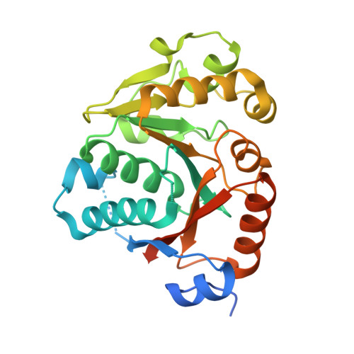
4JLV, 4JMP - PubMed Abstract:
A particular class of tyrosine-kinases sharing no structural similarity with eukaryotic tyrosine-kinases has been evidenced in a large array of bacterial species. These bacterial tyrosine-kinases are able to autophosphorylate on a C-terminal tyrosine-rich motif. Their autophosphorylation has been shown to play a crucial role in the biosynthesis or export of capsular polysaccharide. The analysis of the first crystal structure of the staphylococcal tyrosine kinase CapB2 associated with the activating domain of the transmembrane modulator CapA1 had brought conclusive explanation for both the autophosphorylation and activation processes. In order to explain why CapA1 activates CapB2 more efficiently than its cognate transmembrane modulator CapA2, we solved the crystal structure of CapA2B2 and compared it with the previously published structure of CapA1B2. This structural analysis did not provide the expected clues about the activation discrepancy observed between the two modulators. Staphylococcus aureus also encodes for a CapB2 homologue named CapB1 displaying more than 70% sequence similarity and being surprisingly nearly unable to autophosphorylate. We solved the crystal structure of CapA1B1 and carefully compare it with the structure of CapA1B2. The active sites of both proteins are highly conserved and the biochemical characterization of mutant proteins engineered to test the importance of small structural discrepancies identified between the two structures did not explain the inactivity of CapB1. We thus tested if CapB1 could phosphorylate other protein substrates or hydrolyze ATP. However, no activity could be detected in our in vitro assays. Taken together, these data question about the biological role of the homologous protein pairs CapA1/CapB1 and CapA2/CapB2 and we discuss about several possible interpretations.
- Laboratoire d'Enzymologie et Biochimie Structurales (LEBS), Centre National de la Recherche Scientifique (CNRS), Gif sur Yvette, France.
Organizational Affiliation: