4IZJ

Crystal structure of yellowtail ascites virus VP4 protease with a wild-type active site reveals acyl-enzyme complexes and product complexes.


Experimental Data Snapshot

  • Method: X-RAY DIFFRACTION
  • Resolution: 2.50 Å
  • R-Value Free: 0.244 
  • R-Value Work: 0.178 
  • R-Value Observed: 0.182 

wwPDB Validation   3D Report Full Report


This is version 1.4 of the entry. See complete history


Literature

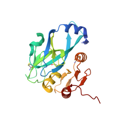
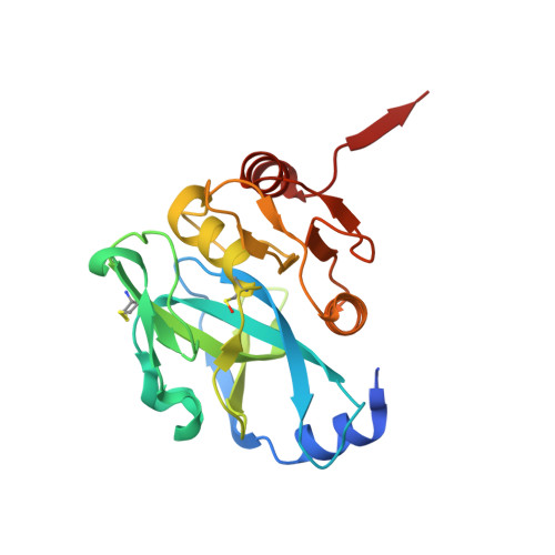
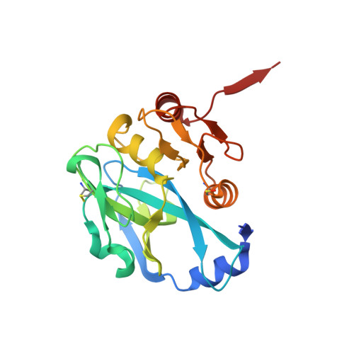
Crystal Structures of Yellowtail Ascites Virus VP4 Protease: TRAPPING AN INTERNAL CLEAVAGE SITE TRANS ACYL-ENZYME COMPLEX IN A NATIVE SER/LYS DYAD ACTIVE SITE.

Chung, I.Y.Paetzel, M.

(2013) J Biol Chem 288: 13068-13081

  • DOI: https://doi.org/10.1074/jbc.M112.386953
  • Primary Citation of Related Structures:  
    4IZJ, 4IZK

  • PubMed Abstract: 

    Yellowtail ascites virus (YAV) is an aquabirnavirus that causes ascites in yellowtail, a fish often used in sushi. Segment A of the YAV genome codes for a polyprotein (pVP2-VP4-VP3), where processing by its own VP4 protease yields the capsid protein precursor pVP2, the ribonucleoprotein-forming VP3, and free VP4. VP4 protease utilizes the rarely observed serine-lysine catalytic dyad mechanism. Here we have confirmed the existence of an internal cleavage site, preceding the VP4/VP3 cleavage site. The resulting C-terminally truncated enzyme (ending at Ala(716)) is active, as shown by a trans full-length VP4 cleavage assay and a fluorometric peptide cleavage assay. We present a crystal structure of a native active site YAV VP4 with the internal cleavage site trapped as trans product complexes and trans acyl-enzyme complexes. The acyl-enzyme complexes confirm directly the role of Ser(633) as the nucleophile. A crystal structure of the lysine general base mutant (K674A) reveals the acyl-enzyme and empty binding site states of VP4, which allows for the observation of structural changes upon substrate or product binding. These snapshots of three different stages in the VP4 protease reaction mechanism will aid in the design of anti-birnavirus compounds, provide insight into previous site-directed mutagenesis results, and contribute to understanding of the serine-lysine dyad protease mechanism. In addition, we have discovered that this protease contains a channel that leads from the enzyme surface (adjacent to the substrate binding groove) to the active site and the deacylating water.


  • Organizational Affiliation

    Department of Molecular Biology and Biochemistry, Simon Fraser University, Burnaby, British Columbia V5A 1S6, Canada.


Macromolecules
Find similar proteins by:  (by identity cutoff)  |  3D Structure
Entity ID: 1
MoleculeChains Sequence LengthOrganismDetailsImage
Yellowtail Ascites Virus (YAV) VP4 protease
A, E
210Yellowtail ascites virus - Y-6Mutation(s): 1 
Gene Names: VIRAL PROTEIN 4 (VP4)
EC: 3.4.21.115
UniProt
Find proteins for P89521 (Yellowtail ascites virus - Y-6)
Explore P89521 
Go to UniProtKB:  P89521
Entity Groups  
Sequence Clusters30% Identity50% Identity70% Identity90% Identity95% Identity100% Identity
UniProt GroupP89521
Sequence Annotations
Expand
  • Reference Sequence
Find similar proteins by:  (by identity cutoff)  |  3D Structure
Entity ID: 2
MoleculeChains Sequence LengthOrganismDetailsImage
Yellowtail Ascites Virus (YAV) VP4 protease
B, C
210Yellowtail ascites virus - Y-6Mutation(s): 1 
Gene Names: VIRAL PROTEIN 4 (VP4)
EC: 3.4.21.115
UniProt
Find proteins for P89521 (Yellowtail ascites virus - Y-6)
Explore P89521 
Go to UniProtKB:  P89521
Entity Groups  
Sequence Clusters30% Identity50% Identity70% Identity90% Identity95% Identity100% Identity
UniProt GroupP89521
Sequence Annotations
Expand
  • Reference Sequence
Find similar proteins by:  (by identity cutoff)  |  3D Structure
Entity ID: 3
MoleculeChains Sequence LengthOrganismDetailsImage
Yellowtail Ascites Virus (YAV) VP4 protease210Yellowtail ascites virus - Y-6Mutation(s): 1 
Gene Names: VIRAL PROTEIN 4 (VP4)
EC: 3.4.21.115
UniProt
Find proteins for P89521 (Yellowtail ascites virus - Y-6)
Explore P89521 
Go to UniProtKB:  P89521
Entity Groups  
Sequence Clusters30% Identity50% Identity70% Identity90% Identity95% Identity100% Identity
UniProt GroupP89521
Sequence Annotations
Expand
  • Reference Sequence
Small Molecules
Ligands 3 Unique
IDChains Name / Formula / InChI Key2D Diagram3D Interactions
GOL
Query on GOL

Download Ideal Coordinates CCD File 
K [auth D]GLYCEROL
C3 H8 O3
PEDCQBHIVMGVHV-UHFFFAOYSA-N
BME
Query on BME

Download Ideal Coordinates CCD File 
F [auth A]
H [auth B]
I [auth B]
J [auth C]
L [auth D]
F [auth A],
H [auth B],
I [auth B],
J [auth C],
L [auth D],
M [auth E]
BETA-MERCAPTOETHANOL
C2 H6 O S
DGVVWUTYPXICAM-UHFFFAOYSA-N
MG
Query on MG

Download Ideal Coordinates CCD File 
G [auth A]MAGNESIUM ION
Mg
JLVVSXFLKOJNIY-UHFFFAOYSA-N
Modified Residues  1 Unique
IDChains TypeFormula2D DiagramParent
CSO
Query on CSO
A, E
L-PEPTIDE LINKINGC3 H7 N O3 SCYS
Experimental Data & Validation

Experimental Data

  • Method: X-RAY DIFFRACTION
  • Resolution: 2.50 Å
  • R-Value Free: 0.244 
  • R-Value Work: 0.178 
  • R-Value Observed: 0.182 
  • Space Group: P 1 21 1
Unit Cell:
Length ( Å )Angle ( ˚ )
a = 41.61α = 90
b = 64.33β = 95.8
c = 187.74γ = 90
Software Package:
Software NamePurpose
CrystalCleardata collection
MOLREPphasing
REFMACrefinement
MOSFLMdata reduction
SCALAdata scaling

Structure Validation

View Full Validation Report


Entry History 

Deposition Data

Revision History  (Full details and data files)

  • Version 1.0: 2013-02-27
    Type: Initial release
  • Version 1.1: 2013-04-03
    Changes: Database references
  • Version 1.2: 2013-05-22
    Changes: Database references
  • Version 1.3: 2023-09-20
    Changes: Data collection, Database references, Derived calculations, Refinement description
  • Version 1.4: 2023-12-06
    Changes: Data collection