


2.0A Crystal Structure of a Glutamate-1-Semialdehyde Aminotransferase from Yersinia pestis CO92
Brunzelle, J.S., Wawrzak, W., Onopriyenko, O., Anderson, W.F., Savchenko, A., Center for Structural Genomics of Infectious DiseasesTo be published.
Experimental Data Snapshot
wwPDB Validation 3D Report Full Report

Entity ID: 1 | |||||
|---|---|---|---|---|---|
| Molecule | Chains | Sequence Length | Organism | Details | Image |
| Glutamate-1-semialdehyde 2,1-aminomutase | 429 | Yersinia pestis Pestoides F | Mutation(s): 0 Gene Names: hemL, YPDSF_2968 EC: 5.4.3.8 | 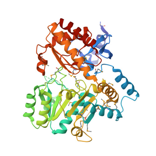 | |
UniProt | |||||
Find proteins for Q8ZBL9 (Yersinia pestis) Explore Q8ZBL9 Go to UniProtKB: Q8ZBL9 | |||||
Entity Groups | |||||
| Sequence Clusters | 30% Identity50% Identity70% Identity90% Identity95% Identity100% Identity | ||||
| UniProt Group | Q8ZBL9 | ||||
Sequence AnnotationsExpand | |||||
| |||||
| Ligands 2 Unique | |||||
|---|---|---|---|---|---|
| ID | Chains | Name / Formula / InChI Key | 2D Diagram | 3D Interactions | |
| NO3 Query on NO3 | C [auth A] | NITRATE ION N O3 NHNBFGGVMKEFGY-UHFFFAOYSA-N |  | ||
| NA Query on NA | B [auth A] | SODIUM ION Na FKNQFGJONOIPTF-UHFFFAOYSA-N |  | ||
| Modified Residues 1 Unique | |||||
|---|---|---|---|---|---|
| ID | Chains | Type | Formula | 2D Diagram | Parent |
| MSE Query on MSE | A | L-PEPTIDE LINKING | C5 H11 N O2 Se |  | MET |
| Length ( Å ) | Angle ( ˚ ) |
|---|---|
| a = 60.54 | α = 90 |
| b = 60.54 | β = 90 |
| c = 182.48 | γ = 120 |
| Software Name | Purpose |
|---|---|
| BLU-MAX | data collection |
| PHENIX | model building |
| BUSTER | refinement |
| XDS | data reduction |
| xia2 | data reduction |
| SCALA | data scaling |
| xia2 | data scaling |
| PHENIX | phasing |


