4WF5

Crystal structure of E.Coli DsbA soaked with compound 4


Experimental Data Snapshot

  • Method: X-RAY DIFFRACTION
  • Resolution: 1.45 Å
  • R-Value Free: 0.196 
  • R-Value Work: 0.160 
  • R-Value Observed: 0.162 

wwPDB Validation   3D Report Full Report


Ligand Structure Quality Assessment 


This is version 1.3 of the entry. See complete history

Re-refinement Note

A newer entry is available that reflects an alternative modeling of the original data: 6BQX


Literature

Application of Fragment-Based Screening to the Design of Inhibitors of Escherichia coli DsbA.

Adams, L.A.Sharma, P.Mohanty, B.Ilyichova, O.V.Mulcair, M.D.Williams, M.L.Gleeson, E.C.Totsika, M.Doak, B.C.Caria, S.Rimmer, K.Horne, J.Shouldice, S.R.Vazirani, M.Headey, S.J.Plumb, B.R.Martin, J.L.Heras, B.Simpson, J.S.Scanlon, M.J.

(2015) Angew Chem Int Ed Engl 54: 2179-2184

  • DOI: https://doi.org/10.1002/anie.201410341
  • Primary Citation of Related Structures:  
    4WET, 4WEY, 4WF4, 4WF5

  • PubMed Abstract: 

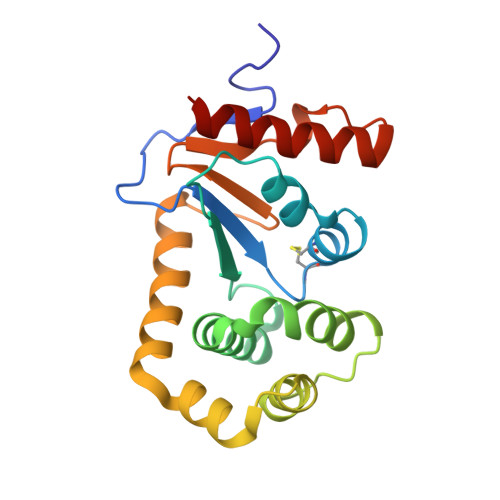
    The thiol-disulfide oxidoreductase enzyme DsbA catalyzes the formation of disulfide bonds in the periplasm of Gram-negative bacteria. DsbA substrates include proteins involved in bacterial virulence. In the absence of DsbA, many of these proteins do not fold correctly, which renders the bacteria avirulent. Thus DsbA is a critical mediator of virulence and inhibitors may act as antivirulence agents. Biophysical screening has been employed to identify fragments that bind to DsbA from Escherichia coli. Elaboration of one of these fragments produced compounds that inhibit DsbA activity in vitro. In cell-based assays, the compounds inhibit bacterial motility, but have no effect on growth in liquid culture, which is consistent with selective inhibition of DsbA. Crystal structures of inhibitors bound to DsbA indicate that they bind adjacent to the active site. Together, the data suggest that DsbA may be amenable to the development of novel antibacterial compounds that act by inhibiting bacterial virulence.


  • Organizational Affiliation

    Medicinal Chemistry, Monash Institute of Pharmaceutical Sciences, Monash University, 381 Royal Parade, Parkville, VIC 3052 (Australia) http://www.pharm.monash.edu.au.


Macromolecules
Find similar proteins by:  (by identity cutoff)  |  3D Structure
Entity ID: 1
MoleculeChains Sequence LengthOrganismDetailsImage
Thiol:disulfide interchange protein
A, B
189Escherichia coli BL21(DE3)Mutation(s): 0 
EC: 1.8.4.2
UniProt
Find proteins for A0A0H2UL03 (Escherichia coli (strain B / BL21-DE3))
Explore A0A0H2UL03 
Go to UniProtKB:  A0A0H2UL03
Entity Groups  
Sequence Clusters30% Identity50% Identity70% Identity90% Identity95% Identity100% Identity
UniProt GroupA0A0H2UL03
Sequence Annotations
Expand
  • Reference Sequence
Small Molecules
Binding Affinity Annotations 
IDSourceBinding Affinity
WF4 Binding MOAD:  4WF5 Kd: 7.77e+5 (nM) from 1 assay(s)
Experimental Data & Validation

Experimental Data

  • Method: X-RAY DIFFRACTION
  • Resolution: 1.45 Å
  • R-Value Free: 0.196 
  • R-Value Work: 0.160 
  • R-Value Observed: 0.162 
  • Space Group: C 1 2 1
Unit Cell:
Length ( Å )Angle ( ˚ )
a = 118.246α = 90
b = 63.401β = 126.19
c = 74.402γ = 90
Software Package:
Software NamePurpose
PHENIXrefinement
Cootmodel building
Blu-Icedata collection
PHASERphasing
SCALAdata reduction
MOSFLMdata processing

Structure Validation

View Full Validation Report


Ligand Structure Quality Assessment 


Entry History & Funding Information

Deposition Data


Funding OrganizationLocationGrant Number
National Health and Medical Research Council (Australia)Australia1009785

Revision History  (Full details and data files)

  • Version 1.0: 2015-01-21
    Type: Initial release
  • Version 1.1: 2015-02-18
    Changes: Database references
  • Version 1.2: 2017-10-18
    Changes: Author supporting evidence, Data collection, Database references, Derived calculations, Other, Source and taxonomy
  • Version 1.3: 2023-11-08
    Changes: Data collection, Database references, Derived calculations, Refinement description