4UEM

UCH-L5 in complex with the RPN13 DEUBAD domain


Experimental Data Snapshot

  • Method: X-RAY DIFFRACTION
  • Resolution: 2.82 Å
  • R-Value Free: 0.281 
  • R-Value Work: 0.232 
  • R-Value Observed: 0.234 

wwPDB Validation   3D Report Full Report


This is version 1.3 of the entry. See complete history


Literature

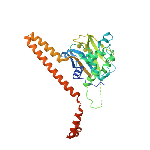
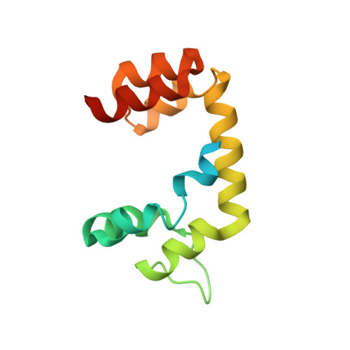
Mechanism of Uch-L5 Activation and Inhibition by Deubad Domains in Rpn13 and Ino80G.

Sahtoe, D.D.Van Dijk, W.J.El Oualid, F.Ekkebus, R.Ovaa, H.Sixma, T.K.

(2015) Mol Cell 57: 887

  • DOI: https://doi.org/10.1016/j.molcel.2014.12.039
  • Primary Citation of Related Structures:  
    4UEL, 4UEM, 4UF5, 4UF6

  • PubMed Abstract: 

    Deubiquitinating enzymes (DUBs) control vital processes in eukaryotes by hydrolyzing ubiquitin adducts. Their activities are tightly regulated, but the mechanisms remain elusive. In particular, the DUB UCH-L5 can be either activated or inhibited by conserved regulatory proteins RPN13 and INO80G, respectively. Here we show how the DEUBAD domain in RPN13 activates UCH-L5 by positioning its C-terminal ULD domain and crossover loop to promote substrate binding and catalysis. The related DEUBAD domain in INO80G inhibits UCH-L5 by exploiting similar structural elements in UCH-L5 to promote a radically different conformation, and employs molecular mimicry to block ubiquitin docking. In this process, large conformational changes create small but highly specific interfaces that mediate activity modulation of UCH-L5 by altering the affinity for substrates. Our results establish how related domains can exploit enzyme conformational plasticity to allosterically regulate DUB activity. These allosteric sites may present novel insights for pharmaceutical intervention in DUB activity.


  • Organizational Affiliation

    Division of Biochemistry and Cancer Genomics Center, The Netherlands Cancer Institute, Plesmanlaan 121, 1066CX Amsterdam, the Netherlands.


Macromolecules
Find similar proteins by:  (by identity cutoff)  |  3D Structure
Entity ID: 1
MoleculeChains Sequence LengthOrganismDetailsImage
UBIQUITIN CARBOXYL-TERMINAL HYDROLASE ISOZYME L5331Homo sapiensMutation(s): 0 
EC: 3.4.19.12
UniProt & NIH Common Fund Data Resources
Find proteins for Q9Y5K5 (Homo sapiens)
Explore Q9Y5K5 
Go to UniProtKB:  Q9Y5K5
PHAROS:  Q9Y5K5
GTEx:  ENSG00000116750 
Entity Groups  
Sequence Clusters30% Identity50% Identity70% Identity90% Identity95% Identity100% Identity
UniProt GroupQ9Y5K5
Sequence Annotations
Expand
  • Reference Sequence
Find similar proteins by:  (by identity cutoff)  |  3D Structure
Entity ID: 2
MoleculeChains Sequence LengthOrganismDetailsImage
PROTEASOMAL UBIQUITIN RECEPTOR ADRM1127Homo sapiensMutation(s): 0 
UniProt & NIH Common Fund Data Resources
Find proteins for Q16186 (Homo sapiens)
Explore Q16186 
Go to UniProtKB:  Q16186
PHAROS:  Q16186
GTEx:  ENSG00000130706 
Entity Groups  
Sequence Clusters30% Identity50% Identity70% Identity90% Identity95% Identity100% Identity
UniProt GroupQ16186
Sequence Annotations
Expand
  • Reference Sequence
Experimental Data & Validation

Experimental Data

  • Method: X-RAY DIFFRACTION
  • Resolution: 2.82 Å
  • R-Value Free: 0.281 
  • R-Value Work: 0.232 
  • R-Value Observed: 0.234 
  • Space Group: P 21 21 21
Unit Cell:
Length ( Å )Angle ( ˚ )
a = 56.596α = 90
b = 97.067β = 90
c = 100.616γ = 90
Software Package:
Software NamePurpose
PHENIXrefinement
Aimlessdata scaling
PHASERphasing

Structure Validation

View Full Validation Report


Entry History 

Deposition Data

Revision History  (Full details and data files)

  • Version 1.0: 2015-03-04
    Type: Initial release
  • Version 1.1: 2015-03-25
    Changes: Database references
  • Version 1.2: 2019-04-03
    Changes: Data collection, Experimental preparation, Other
  • Version 1.3: 2023-12-20
    Changes: Data collection, Database references, Other, Refinement description