4R0Y

Structure of Maltose-binding Protein Fusion with the C-terminal GH1 domain of Guanylate Kinase-associated Protein from Rattus norvegicus


Experimental Data Snapshot

  • Method: X-RAY DIFFRACTION
  • Resolution: 2.00 Å
  • R-Value Free: 0.281 
  • R-Value Work: 0.243 
  • R-Value Observed: 0.243 

wwPDB Validation   3D Report Full Report


This is version 1.3 of the entry. See complete history


Literature

Structure of the GH1 domain of guanylate kinase-associated protein from Rattus norvegicus.

Tong, J.Yang, H.Eom, S.H.Chun, C.Im, Y.J.

(2014) Biochem Biophys Res Commun 452: 130-135

  • DOI: https://doi.org/10.1016/j.bbrc.2014.08.073
  • Primary Citation of Related Structures:  
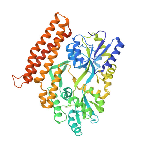
    4R0Y

  • PubMed Abstract: 

    Guanylate-kinase-associated protein (GKAP) is a scaffolding protein that links NMDA receptor-PSD-95 to Shank-Homer complexes by protein-protein interactions at the synaptic junction. GKAP family proteins are characterized by the presence of a C-terminal conserved GKAP homology domain 1 (GH1) of unknown structure and function. In this study, crystal structure of the GH1 domain of GKAP from Rattus norvegicus was determined in fusion with an N-terminal maltose-binding protein at 2.0 Å resolution. The structure of GKAP GH1 displays a three-helix bundle connected by short flexible loops. The predicted helix α4 which was not visible in the crystal structure associates weakly with the helix α3 suggesting dynamic nature of the GH1 domain. The strict conservation of GH1 domain across GKAP family members and the lack of a catalytic active site required for enzyme activity imply that the GH1 domain might serve as a protein-protein interaction module for the synaptic protein clustering.


  • Organizational Affiliation

    College of Pharmacy, Chonnam National University, Gwangju 500-757, South Korea.


Macromolecules
Find similar proteins by:  (by identity cutoff)  |  3D Structure
Entity ID: 1
MoleculeChains Sequence LengthOrganismDetailsImage
Maltose-binding periplasmic protein, Disks large-associated protein 1
A, B
501Escherichia coli K-12Rattus norvegicusMutation(s): 0 
UniProt
Find proteins for P0AEX9 (Escherichia coli (strain K12))
Explore P0AEX9 
Go to UniProtKB:  P0AEX9
Find proteins for P97836 (Rattus norvegicus)
Explore P97836 
Go to UniProtKB:  P97836
Entity Groups  
Sequence Clusters30% Identity50% Identity70% Identity90% Identity95% Identity100% Identity
UniProt GroupsP97836P0AEX9
Sequence Annotations
Expand
  • Reference Sequence
Experimental Data & Validation

Experimental Data

  • Method: X-RAY DIFFRACTION
  • Resolution: 2.00 Å
  • R-Value Free: 0.281 
  • R-Value Work: 0.243 
  • R-Value Observed: 0.243 
  • Space Group: P 21 21 2
Unit Cell:
Length ( Å )Angle ( ˚ )
a = 99.11α = 90
b = 158.676β = 90
c = 65.494γ = 90
Software Package:
Software NamePurpose
ADSCdata collection
MOLREPphasing
CNSrefinement
HKL-2000data reduction
HKL-2000data scaling

Structure Validation

View Full Validation Report


Entry History 

Deposition Data

Revision History  (Full details and data files)

  • Version 1.0: 2014-09-10
    Type: Initial release
  • Version 1.1: 2017-08-16
    Changes: Refinement description, Source and taxonomy
  • Version 1.2: 2022-08-24
    Changes: Database references
  • Version 1.3: 2023-11-08
    Changes: Data collection, Refinement description