


Crystal structures of E.Coli glutaredoxin 2 with glutathione and of a mutant C9S/C12S without glutathione
Ye, J., Venkadesh, S., Rosen, B.P.To be published.
Experimental Data Snapshot
wwPDB Validation 3D Report Full Report

Entity ID: 1 | |||||
|---|---|---|---|---|---|
| Molecule | Chains | Sequence Length | Organism | Details | Image |
| Glutaredoxin-2 | 216 | Escherichia coli K-12 | Mutation(s): 2 Gene Names: grxB, b1064, JW1051 | 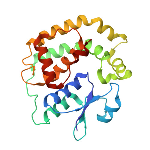 | |
UniProt | |||||
Find proteins for P0AC59 (Escherichia coli (strain K12)) Explore P0AC59 Go to UniProtKB: P0AC59 | |||||
Entity Groups | |||||
| Sequence Clusters | 30% Identity50% Identity70% Identity90% Identity95% Identity100% Identity | ||||
| UniProt Group | P0AC59 | ||||
Sequence AnnotationsExpand | |||||
| |||||
| Ligands 1 Unique | |||||
|---|---|---|---|---|---|
| ID | Chains | Name / Formula / InChI Key | 2D Diagram | 3D Interactions | |
| TRS Query on TRS | B [auth A] | 2-AMINO-2-HYDROXYMETHYL-PROPANE-1,3-DIOL C4 H12 N O3 LENZDBCJOHFCAS-UHFFFAOYSA-O |  | ||
| Length ( Å ) | Angle ( ˚ ) |
|---|---|
| a = 28.16 | α = 90 |
| b = 78.65 | β = 90 |
| c = 89.16 | γ = 90 |
| Software Name | Purpose |
|---|---|
| CrystalClear | data collection |
| AMoRE | phasing |
| PHENIX | refinement |
| CrystalClear | data reduction |
| CrystalClear | data scaling |


