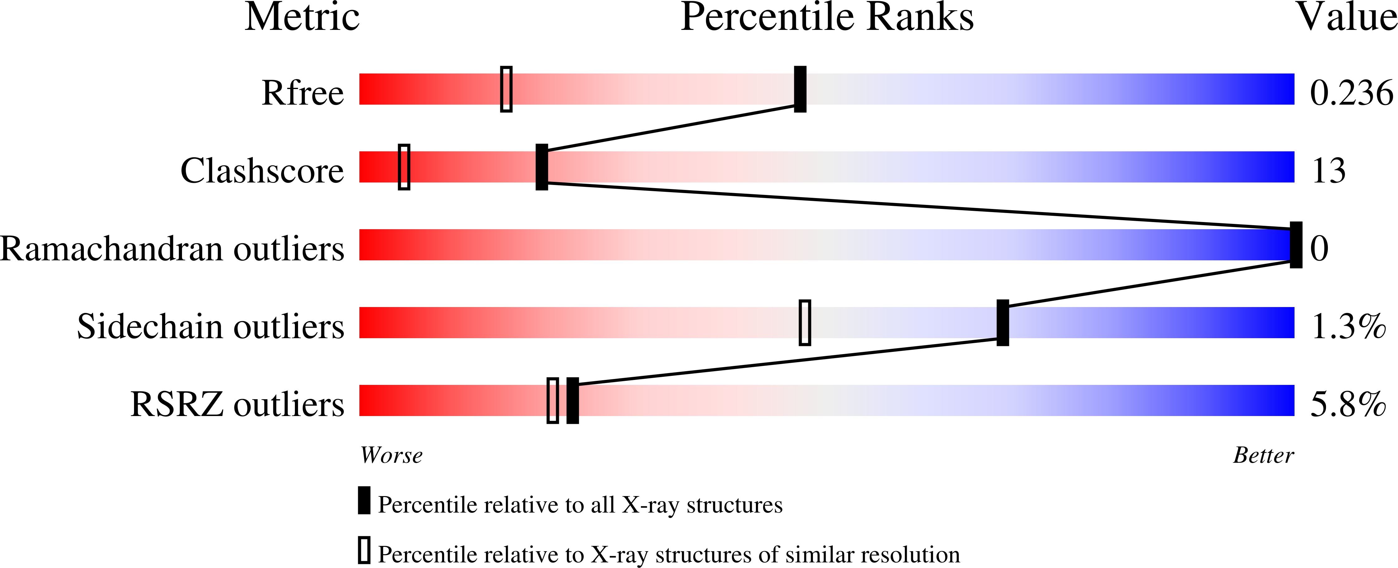
4J7E

The 1.63A crystal structure of humanized Xenopus MDM2 with a nutlin fragment, RO5524529


Experimental Data Snapshot

  • Method: X-RAY DIFFRACTION
  • Resolution: 1.63 Å
  • R-Value Free: 0.237 
  • R-Value Work: 0.222 
  • R-Value Observed: 0.223 

wwPDB Validation   3D Report Full Report


Ligand Structure Quality Assessment 


This is version 1.2 of the entry. See complete history


Literature

Deconstruction of a nutlin: dissecting the binding determinants of a potent protein-protein interaction inhibitor.

Fry, D.C.Wartchow, C.Graves, B.Janson, C.Lukacs, C.Kammlott, U.Belunis, C.Palme, S.Klein, C.Vu, B.

(2013) ACS Med Chem Lett 4: 660-665

  • DOI: https://doi.org/10.1021/ml400062c
  • Primary Citation of Related Structures:  
    4J74, 4J7D, 4J7E

  • PubMed Abstract: 

    Protein-protein interaction (PPI) systems represent a rich potential source of targets for drug discovery, but historically have proven to be difficult, particularly in the lead identification stage. Application of the fragment-based approach may help toward success with this target class. To provide an example toward understanding the potential issues associated with such an application, we have deconstructed one of the best established protein-protein inhibitors, the Nutlin series that inhibits the interaction between MDM2 and p53, into fragments, and surveyed the resulting binding properties using heteronuclear single quantum coherence nuclear magnetic resonance (HSQC NMR), surface plasmon resonance (SPR), and X-ray crystallography. We report the relative contributions toward binding affinity for each of the key substituents of the Nutlin molecule and show that this series could hypothetically have been discovered via a fragment approach. We find that the smallest fragment of Nutlin that retains binding accesses two subpockets of MDM2 and has a molecular weight at the high end of the range that normally defines fragments.


  • Organizational Affiliation

    Roche Research Center , 340 Kingsland Street, Nutley, New Jersey 07110, United States.


Macromolecules
Find similar proteins by:  (by identity cutoff)  |  3D Structure
Entity ID: 1
MoleculeChains Sequence LengthOrganismDetailsImage
E3 ubiquitin-protein ligase Mdm286Xenopus laevisMutation(s): 3 
Gene Names: mdm2
EC: 6.3.2
UniProt
Find proteins for P56273 (Xenopus laevis)
Explore P56273 
Go to UniProtKB:  P56273
Entity Groups  
Sequence Clusters30% Identity50% Identity70% Identity90% Identity95% Identity100% Identity
UniProt GroupP56273
Sequence Annotations
Expand
  • Reference Sequence
Small Molecules
Ligands 2 Unique
IDChains Name / Formula / InChI Key2D Diagram3D Interactions
I29
Query on I29

Download Ideal Coordinates CCD File 
B [auth A][(4S,5R)-4,5-bis(4-chlorophenyl)-2,4,5-trimethyl-4,5-dihydro-1H-imidazol-1-yl]{4-[3-(methylsulfonyl)propyl]piperazin-1-yl}methanone
C27 H34 Cl2 N4 O3 S
MCLBNCWBALNIBY-RRPNLBNLSA-N
SO4
Query on SO4

Download Ideal Coordinates CCD File 
C [auth A]SULFATE ION
O4 S
QAOWNCQODCNURD-UHFFFAOYSA-L
Binding Affinity Annotations 
IDSourceBinding Affinity
I29 Binding MOAD:  4J7E Kd: 1.40e+4 (nM) from 1 assay(s)
PDBBind:  4J7E Kd: 1.40e+4 (nM) from 1 assay(s)
Experimental Data & Validation

Experimental Data

  • Method: X-RAY DIFFRACTION
  • Resolution: 1.63 Å
  • R-Value Free: 0.237 
  • R-Value Work: 0.222 
  • R-Value Observed: 0.223 
  • Space Group: C 2 2 21
Unit Cell:
Length ( Å )Angle ( ˚ )
a = 43.29α = 90
b = 68.612β = 90
c = 67.349γ = 90
Software Package:
Software NamePurpose
HKL-3000data collection
MOLREPphasing
CNXrefinement
HKL-3000data reduction
SCALAdata scaling

Structure Validation

View Full Validation Report


Ligand Structure Quality Assessment 


Entry History 

Deposition Data

Revision History  (Full details and data files)

  • Version 1.0: 2013-08-07
    Type: Initial release
  • Version 1.1: 2014-09-24
    Changes: Database references
  • Version 1.2: 2024-02-28
    Changes: Data collection, Database references, Derived calculations