4IRL

X-ray structure of the CARD domain of zebrafish GBP-NLRP1 like protein


Experimental Data Snapshot

  • Method: X-RAY DIFFRACTION
  • Resolution: 1.47 Å
  • R-Value Free: 0.224 
  • R-Value Work: 0.188 
  • R-Value Observed: 0.190 

wwPDB Validation   3D Report Full Report


This is version 2.1 of the entry. See complete history


Literature

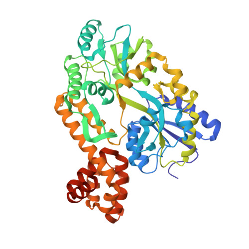
Structure of the caspase-recruitment domain from a zebrafish guanylate-binding protein.

Jin, T.Huang, M.Smith, P.Jiang, J.Xiao, T.S.

(2013) Acta Crystallogr Sect F Struct Biol Cryst Commun 69: 855-860

  • DOI: https://doi.org/10.1107/S1744309113015558
  • Primary Citation of Related Structures:  
    4IRL

  • PubMed Abstract: 

    The caspase-recruitment domain (CARD) mediates homotypic protein-protein interactions that assemble large oligomeric signaling complexes such as the inflammasomes during innate immune responses. Structural studies of the mammalian CARDs demonstrate that their six-helix bundle folds belong to the death-domain superfamily, whereas such studies have not been reported for other organisms. Here, the zebrafish interferon-induced guanylate-binding protein 1 (zIGBP1) was identified that contains an N-terminal GTPase domain and a helical domain typical of the mammalian guanylate-binding proteins, followed by a FIIND domain and a C-terminal CARD similar to the mammalian inflammasome proteins NLRP1 and CARD8. The structure of the zIGBP1 CARD as a fusion with maltose-binding protein was determined at 1.47 Å resolution. This revealed a six-helix bundle fold similar to the NLRP1 CARD structure with the bent α1 helix typical of all known CARD structures. The zIGBP1 CARD surface contains a positively charged patch near its α1 and α4 helices and a negatively charged patch near its α2, α3 and α5 helices, which may mediate its interaction with partner domains. Further studies using binding assays and other analyses will be required in order to address the physiological function(s) of this zebrafish protein.


  • Organizational Affiliation

    Structural Immunobiology Unit, Laboratory of Immunology, National Institute of Allergy and Infectious Diseases, National Institutes of Health, 4 Memorial Drive, Building 4, Room 228, Bethesda, MD 20892-0430, USA.


Macromolecules
Find similar proteins by:  (by identity cutoff)  |  3D Structure
Entity ID: 1
MoleculeChains Sequence LengthOrganismDetailsImage
Maltose-binding periplasmic protein, Novel protein similar to vertebrate guanylate binding protein family
A, B, C
476Escherichia coli K-12Danio rerio
This entity is chimeric
Mutation(s): 0 
Gene Names: b4034GBP-NLRP1JW3994malECH211-195H23.5-001gbp3
UniProt
Find proteins for P0AEX9 (Escherichia coli (strain K12))
Explore P0AEX9 
Go to UniProtKB:  P0AEX9
Find proteins for B0V1H4 (Danio rerio)
Explore B0V1H4 
Go to UniProtKB:  B0V1H4
Entity Groups  
Sequence Clusters30% Identity50% Identity70% Identity90% Identity95% Identity100% Identity
UniProt GroupsB0V1H4P0AEX9
Sequence Annotations
Expand
  • Reference Sequence
Oligosaccharides

Help

Entity ID: 2
MoleculeChains Length2D Diagram Glycosylation3D Interactions
alpha-D-glucopyranose-(1-4)-alpha-D-glucopyranose-(1-4)-alpha-D-glucopyranose-(1-4)-alpha-D-glucopyranose
D, E, F
4N/A
Glycosylation Resources
GlyTouCan:  G87171PZ
GlyCosmos:  G87171PZ
GlyGen:  G87171PZ
Small Molecules
Ligands 5 Unique
IDChains Name / Formula / InChI Key2D Diagram3D Interactions
PEG
Query on PEG

Download Ideal Coordinates CCD File 
T [auth C]DI(HYDROXYETHYL)ETHER
C4 H10 O3
MTHSVFCYNBDYFN-UHFFFAOYSA-N
MLI
Query on MLI

Download Ideal Coordinates CCD File 
G [auth A],
K [auth B],
R [auth C]
MALONATE ION
C3 H2 O4
OFOBLEOULBTSOW-UHFFFAOYSA-L
EDO
Query on EDO

Download Ideal Coordinates CCD File 
H [auth A],
L [auth B],
M [auth B],
N [auth B],
S [auth C]
1,2-ETHANEDIOL
C2 H6 O2
LYCAIKOWRPUZTN-UHFFFAOYSA-N
ACT
Query on ACT

Download Ideal Coordinates CCD File 
I [auth A],
O [auth B],
U [auth C]
ACETATE ION
C2 H3 O2
QTBSBXVTEAMEQO-UHFFFAOYSA-M
NA
Query on NA

Download Ideal Coordinates CCD File 
J [auth A],
P [auth B],
Q [auth B],
V [auth C]
SODIUM ION
Na
FKNQFGJONOIPTF-UHFFFAOYSA-N
Biologically Interesting Molecules (External Reference) 1 Unique
Experimental Data & Validation

Experimental Data

  • Method: X-RAY DIFFRACTION
  • Resolution: 1.47 Å
  • R-Value Free: 0.224 
  • R-Value Work: 0.188 
  • R-Value Observed: 0.190 
  • Space Group: P 21 21 21
Unit Cell:
Length ( Å )Angle ( ˚ )
a = 65.749α = 90
b = 124.75β = 90
c = 178.044γ = 90
Software Package:
Software NamePurpose
Blu-Icedata collection
PHASERphasing
PHENIXrefinement
XDSdata reduction
XDSdata scaling

Structure Validation

View Full Validation Report


Entry History 

Deposition Data

Revision History  (Full details and data files)

  • Version 1.0: 2013-08-07
    Type: Initial release
  • Version 1.1: 2013-08-21
    Changes: Database references
  • Version 1.2: 2014-11-26
    Changes: Refinement description
  • Version 1.3: 2017-08-02
    Changes: Refinement description, Source and taxonomy
  • Version 1.4: 2017-11-15
    Changes: Refinement description
  • Version 2.0: 2020-07-29
    Type: Remediation
    Reason: Carbohydrate remediation
    Changes: Atomic model, Data collection, Database references, Derived calculations, Non-polymer description, Structure summary
  • Version 2.1: 2024-02-28
    Changes: Data collection, Database references, Structure summary