4IR7

Crystal Structure of Mtb FadD10 in Complex with Dodecanoyl-AMP


Experimental Data Snapshot

  • Method: X-RAY DIFFRACTION
  • Resolution: 2.80 Å
  • R-Value Free: 0.280 
  • R-Value Work: 0.226 
  • R-Value Observed: 0.229 

wwPDB Validation   3D Report Full Report


Ligand Structure Quality Assessment 


This is version 1.3 of the entry. See complete history


Literature

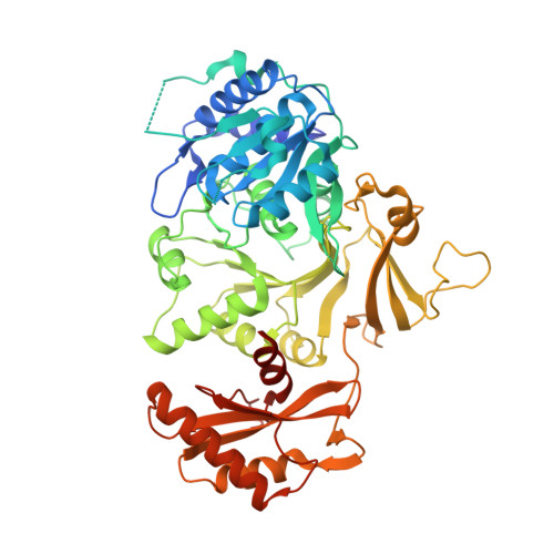
Structures of Mycobacterium tuberculosis FadD10 protein reveal a new type of adenylate-forming enzyme.

Liu, Z.Ioerger, T.R.Wang, F.Sacchettini, J.C.

(2013) J Biol Chem 288: 18473-18483

  • DOI: https://doi.org/10.1074/jbc.M113.466912
  • Primary Citation of Related Structures:  
    4IR7, 4ISB

  • PubMed Abstract: 

    Mycobacterium tuberculosis has a group of 34 FadD proteins that belong to the adenylate-forming superfamily. They are classified as either fatty acyl-AMP ligases (FAALs) or fatty acyl-CoA ligases based on sequence analysis. FadD10, involved in the synthesis of a virulence-related lipopeptide, was mis-annotated as a fatty acyl-CoA ligase; however, it is in fact a FAAL that transfers fatty acids to an acyl carrier protein (Rv0100). In this study, we have determined the structures of FadD10 in both the apo-form and the complexed form with dodecanoyl-AMP, where we see for the first time an adenylate-forming enzyme that does not adopt a closed conformation for catalysis. Indeed, this novel conformation of FadD10, facilitated by its unique inter-domain and intermolecular interactions, is critical for the enzyme to carry out the acyl transfer onto Rv0100 rather than coenzyme A. This contradicts the existing model of FAALs that rely on an insertion motif for the acyltransferase specificity and thus makes FadD10 a new type of FAAL. We have also characterized the fatty acid preference of FadD10 through biological and structural analyses, and the data indicate long chain saturated fatty acids as the biological substrates of the enzyme.


  • Organizational Affiliation

    Department of Chemistry, Texas A&M University, College Station, Texas 77842-3012, USA.


Macromolecules
Find similar proteins by:  (by identity cutoff)  |  3D Structure
Entity ID: 1
MoleculeChains Sequence LengthOrganismDetailsImage
Long chain fatty acid CoA ligase FadD10541Mycobacterium tuberculosis H37RvMutation(s): 0 
Gene Names: Rv0099
EC: 2.3.1.86
Entity Groups  
Sequence Clusters30% Identity50% Identity70% Identity90% Identity95% Identity100% Identity
Sequence Annotations
Expand
  • Reference Sequence
Small Molecules
Ligands 2 Unique
IDChains Name / Formula / InChI Key2D Diagram3D Interactions
1ZZ
Query on 1ZZ

Download Ideal Coordinates CCD File 
B [auth A]5'-O-[(S)-(dodecanoyloxy)(hydroxy)phosphoryl]adenosine
C22 H36 N5 O8 P
IKBWVSPLSBIYSK-CIVUBGFFSA-N
MG
Query on MG

Download Ideal Coordinates CCD File 
C [auth A]MAGNESIUM ION
Mg
JLVVSXFLKOJNIY-UHFFFAOYSA-N
Experimental Data & Validation

Experimental Data

  • Method: X-RAY DIFFRACTION
  • Resolution: 2.80 Å
  • R-Value Free: 0.280 
  • R-Value Work: 0.226 
  • R-Value Observed: 0.229 
  • Space Group: P 31 2 1
Unit Cell:
Length ( Å )Angle ( ˚ )
a = 138.212α = 90
b = 138.212β = 90
c = 82.582γ = 120
Software Package:
Software NamePurpose
DENZOdata reduction
SCALEPACKdata scaling
PHENIXrefinement
PDB_EXTRACTdata extraction
CrystalCleardata collection
HKL-2000data reduction
AMoREphasing

Structure Validation

View Full Validation Report


Ligand Structure Quality Assessment 


Entry History 

Deposition Data

Revision History  (Full details and data files)

  • Version 1.0: 2013-05-08
    Type: Initial release
  • Version 1.1: 2014-08-20
    Changes: Database references
  • Version 1.2: 2017-11-15
    Changes: Refinement description
  • Version 1.3: 2024-02-28
    Changes: Data collection, Database references, Derived calculations