Identification of an essential active-site residue in the alpha-D-phosphohexomutase enzyme superfamily.
Lee, Y., Mehra-Chaudhary, R., Furdui, C., Beamer, L.J.(2013) FEBS J 280: 2622-2632
- PubMed: 23517223
- DOI: https://doi.org/10.1111/febs.12249
- Primary Citation of Related Structures:
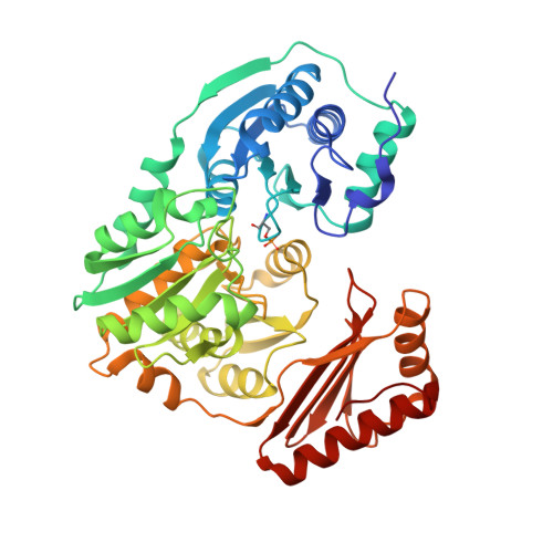
4IL8 - PubMed Abstract:
Enzymes in the α-d-phosphohexomutase superfamily catalyze the conversion of 1-phosphosugars to their 6-phospho counterparts. Their phosphoryl transfer reaction has long been proposed to require general acid-base catalysts, but candidate residues for these key roles have not been identified. In this study, we show through mutagenesis and kinetic studies that a histidine (His329) in the active site is critical for enzyme activity in a well-studied member of the superfamily, phosphomannomutase/phosphoglucomutase from Pseudomonas aeruginosa. Crystallographic characterization of an H329A mutant protein showed no significant changes from the wild-type enzyme, excluding structural disruption as the source of its compromised activity. Mutation of the structurally analogous lysine residue in a related protein, phosphoglucomutase from Salmonella typhimurium, also results in significant catalytic impairment. Analyses of protein-ligand complexes of the P. aeruginosa enzyme show that His329 is appropriately positioned to abstract a proton from the O1/O6 hydroxyl of the phosphosugar substrates, and thus may serve as the general base in the reaction. Histidine is strongly conserved at this position in many proteins in the superfamily, and lysine is also often conserved at a structurally corresponding position, particularly in the phosphoglucomutase enzyme sub-group. These studies shed light on the mechanism of this important enzyme superfamily, and may facilitate the design of mechanism-based inhibitors. Structural data have been deposited in the Protein Data Bank with accession number 4IL8.
- Chemistry Department, University of Missouri, Columbia, MO 65211, USA.
Organizational Affiliation: