4IEM

Human apurinic/apyrimidinic endonuclease (APE1) with product DNA and Mg2+


Experimental Data Snapshot

  • Method: X-RAY DIFFRACTION
  • Resolution: 2.39 Å
  • R-Value Free: 0.246 
  • R-Value Work: 0.208 
  • R-Value Observed: 0.210 

wwPDB Validation   3D Report Full Report


This is version 1.4 of the entry. See complete history


Literature

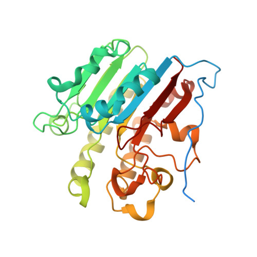
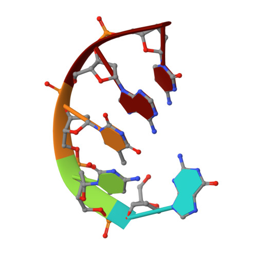
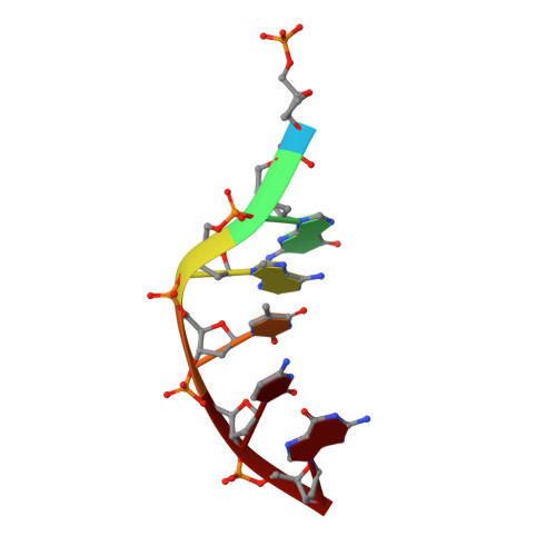
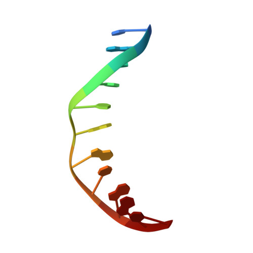
Conserved Structural Chemistry for Incision Activity in Structurally Non-homologous Apurinic/Apyrimidinic Endonuclease APE1 and Endonuclease IV DNA Repair Enzymes.

Tsutakawa, S.E.Shin, D.S.Mol, C.D.Izumi, T.Arvai, A.S.Mantha, A.K.Szczesny, B.Ivanov, I.N.Hosfield, D.J.Maiti, B.Pique, M.E.Frankel, K.A.Hitomi, K.Cunningham, R.P.Mitra, S.Tainer, J.A.

(2013) J Biol Chem 288: 8445-8455

  • DOI: https://doi.org/10.1074/jbc.M112.422774
  • Primary Citation of Related Structures:  
    4HNO, 4IEM

  • PubMed Abstract: 

    Non-coding apurinic/apyrimidinic (AP) sites in DNA form spontaneously and as DNA base excision repair intermediates are the most common toxic and mutagenic in vivo DNA lesion. For repair, AP sites must be processed by 5' AP endonucleases in initial stages of base repair. Human APE1 and bacterial Nfo represent the two conserved 5' AP endonuclease families in the biosphere; they both recognize AP sites and incise the phosphodiester backbone 5' to the lesion, yet they lack similar structures and metal ion requirements. Here, we determined and analyzed crystal structures of a 2.4 Å resolution APE1-DNA product complex with Mg(2+) and a 0.92 Å Nfo with three metal ions. Structural and biochemical comparisons of these two evolutionarily distinct enzymes characterize key APE1 catalytic residues that are potentially functionally similar to Nfo active site components, as further tested and supported by computational analyses. We observe a magnesium-water cluster in the APE1 active site, with only Glu-96 forming the direct protein coordination to the Mg(2+). Despite differences in structure and metal requirements of APE1 and Nfo, comparison of their active site structures surprisingly reveals strong geometric conservation of the catalytic reaction, with APE1 catalytic side chains positioned analogously to Nfo metal positions, suggesting surprising functional equivalence between Nfo metal ions and APE1 residues. The finding that APE1 residues are positioned to substitute for Nfo metal ions is supported by the impact of mutations on activity. Collectively, the results illuminate the activities of residues, metal ions, and active site features for abasic site endonucleases.


  • Organizational Affiliation

    Lawrence Berkeley National Laboratory, Berkeley, California 94720.


Macromolecules

Find similar proteins by:  (by identity cutoff)  |  3D Structure
Entity ID: 1
MoleculeChains Sequence LengthOrganismDetailsImage
DNA-(apurinic or apyrimidinic site) lyaseA,
E [auth B],
I [auth C],
M [auth D]
317Homo sapiensMutation(s): 0 
Gene Names: APEAPE1APEXAPEX1APXHAP1REF1
EC: 3.1 (PDB Primary Data), 4.2.99.18 (PDB Primary Data)
UniProt & NIH Common Fund Data Resources
Find proteins for P27695 (Homo sapiens)
Explore P27695 
Go to UniProtKB:  P27695
PHAROS:  P27695
GTEx:  ENSG00000100823 
Entity Groups  
Sequence Clusters30% Identity50% Identity70% Identity90% Identity95% Identity100% Identity
UniProt GroupP27695
Sequence Annotations
Expand
  • Reference Sequence

Find similar nucleic acids by:  Sequence   |   3D Structure  

Entity ID: 2
MoleculeChains LengthOrganismImage
DNA (5'-D(*GP*CP*TP*AP*C)-3')B [auth E],
F [auth H],
J [auth K],
N
5synthetic construct
Sequence Annotations
Expand
  • Reference Sequence

Find similar nucleic acids by:  Sequence   |   3D Structure  

Entity ID: 3
MoleculeChains LengthOrganismImage
DNA (5'-D(P*(3DR)P*GP*AP*TP*CP*G)-3')C [auth F],
G [auth I],
K [auth L],
O
6synthetic construct
Sequence Annotations
Expand
  • Reference Sequence

Find similar nucleic acids by:  Sequence   |   3D Structure  

Entity ID: 4
MoleculeChains LengthOrganismImage
DNA (5'-D(*CP*GP*AP*TP*CP*GP*GP*TP*AP*GP*C)-3')D [auth G],
H [auth J],
L [auth M],
P
11synthetic construct
Sequence Annotations
Expand
  • Reference Sequence
Small Molecules
Ligands 2 Unique
IDChains Name / Formula / InChI Key2D Diagram3D Interactions
MG
Query on MG

Download Ideal Coordinates CCD File 
BA [auth C]
CA [auth C]
DA [auth D]
Q [auth A]
R [auth A]
BA [auth C],
CA [auth C],
DA [auth D],
Q [auth A],
R [auth A],
U [auth A],
X [auth B]
MAGNESIUM ION
Mg
JLVVSXFLKOJNIY-UHFFFAOYSA-N
NA
Query on NA

Download Ideal Coordinates CCD File 
AA [auth B]
EA [auth D]
FA [auth N]
S [auth A]
T [auth A]
AA [auth B],
EA [auth D],
FA [auth N],
S [auth A],
T [auth A],
V [auth E],
W [auth F],
Y [auth B],
Z [auth B]
SODIUM ION
Na
FKNQFGJONOIPTF-UHFFFAOYSA-N
Experimental Data & Validation

Experimental Data

  • Method: X-RAY DIFFRACTION
  • Resolution: 2.39 Å
  • R-Value Free: 0.246 
  • R-Value Work: 0.208 
  • R-Value Observed: 0.210 
  • Space Group: P 1 21 1
Unit Cell:
Length ( Å )Angle ( ˚ )
a = 104.639α = 90
b = 74.09β = 111.98
c = 112.138γ = 90
Software Package:
Software NamePurpose
SCALEPACKdata scaling
AMoREphasing
PHENIXrefinement
PDB_EXTRACTdata extraction
DENZOdata reduction

Structure Validation

View Full Validation Report


Entry History 

Deposition Data

Revision History  (Full details and data files)

  • Version 1.0: 2013-01-23
    Type: Initial release
  • Version 1.1: 2013-02-13
    Changes: Database references, Derived calculations, Other
  • Version 1.2: 2013-04-10
    Changes: Database references
  • Version 1.3: 2017-11-15
    Changes: Refinement description
  • Version 1.4: 2024-02-28
    Changes: Data collection, Database references, Derived calculations