4CJX

The crystal structure of Trypanosoma brucei N5, N10- methylenetetrahydrofolate dehydrogenase-cyclohydrolase (FolD) complexed with NADP cofactor and inhibitor


Experimental Data Snapshot

  • Method: X-RAY DIFFRACTION
  • Resolution: 2.05 Å
  • R-Value Free: 0.228 
  • R-Value Work: 0.180 
  • R-Value Observed: 0.183 

wwPDB Validation   3D Report Full Report


Ligand Structure Quality Assessment 


This is version 1.2 of the entry. See complete history


Literature

Characterization of 2,4-Diamino-6-Oxo-1,6-Dihydropyrimidin-5-Yl Ureido Based Inhibitors of Trypanosoma Brucei Fold and Testing for Antiparasitic Activity.

Eadsforth, T.C.Pinto, A.Luciani, R.Tamborini, L.Cullia, G.De Micheli, C.Marinelli, L.Cosconati, S.Novellino, E.Lo Presti, L.Cordeiro Da Silva, A.Conti, P.Hunter, W.N.Costi, M.P.

(2015) J Med Chem 58: 7938

  • DOI: https://doi.org/10.1021/acs.jmedchem.5b00687
  • Primary Citation of Related Structures:  
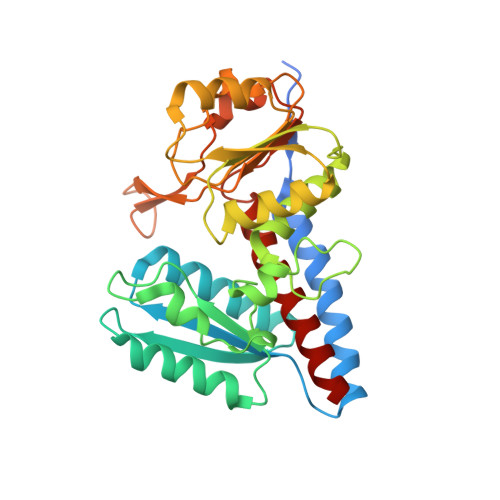
    4CJX

  • PubMed Abstract: 

    The bifunctional enzyme N(5),N(10)-methylenetetrahydrofolate dehydrogenase/cyclo hydrolase (FolD) is essential for growth in Trypanosomatidae. We sought to develop inhibitors of Trypanosoma brucei FolD (TbFolD) as potential antiparasitic agents. Compound 2 was synthesized, and the molecular structure was unequivocally assigned through X-ray crystallography of the intermediate compound 3. Compound 2 showed an IC50 of 2.2 μM, against TbFolD and displayed antiparasitic activity against T. brucei (IC50 49 μM). Using compound 2, we were able to obtain the first X-ray structure of TbFolD in the presence of NADP(+) and the inhibitor, which then guided the rational design of a new series of potent TbFolD inhibitors.


  • Organizational Affiliation

    Division of Biological Chemistry and Drug Discovery, College of Life Sciences, University of Dundee , Dow Street, Dundee DD1 5EH, U.K.


Macromolecules
Find similar proteins by:  (by identity cutoff)  |  3D Structure
Entity ID: 1
MoleculeChains Sequence LengthOrganismDetailsImage
C-1-TETRAHYDROFOLATE SYNTHASE, CYTOPLASMIC, PUTATIVE
A, B
319Trypanosoma brucei brucei TREU927Mutation(s): 0 
EC: 1.5.1.5
UniProt
Find proteins for Q57WP0 (Trypanosoma brucei brucei (strain 927/4 GUTat10.1))
Explore Q57WP0 
Go to UniProtKB:  Q57WP0
Entity Groups  
Sequence Clusters30% Identity50% Identity70% Identity90% Identity95% Identity100% Identity
UniProt GroupQ57WP0
Sequence Annotations
Expand
  • Reference Sequence
Small Molecules
Ligands 4 Unique
IDChains Name / Formula / InChI Key2D Diagram3D Interactions
NAP
Query on NAP

Download Ideal Coordinates CCD File 
F [auth A]NADP NICOTINAMIDE-ADENINE-DINUCLEOTIDE PHOSPHATE
C21 H28 N7 O17 P3
XJLXINKUBYWONI-NNYOXOHSSA-N
9L9
Query on 9L9

Download Ideal Coordinates CCD File 
E [auth A],
G [auth B]
(2S)-2-[[4-[[2,4-bis(azanyl)-6-oxidanylidene-1H-pyrimidin-5-yl]carbamoylamino]phenyl]carbonylamino]pentanedioic acid
C17 H19 N7 O7
WBQDVZMETQWVQD-VIFPVBQESA-N
PEG
Query on PEG

Download Ideal Coordinates CCD File 
D [auth A]DI(HYDROXYETHYL)ETHER
C4 H10 O3
MTHSVFCYNBDYFN-UHFFFAOYSA-N
GOL
Query on GOL

Download Ideal Coordinates CCD File 
C [auth A]GLYCEROL
C3 H8 O3
PEDCQBHIVMGVHV-UHFFFAOYSA-N
Experimental Data & Validation

Experimental Data

  • Method: X-RAY DIFFRACTION
  • Resolution: 2.05 Å
  • R-Value Free: 0.228 
  • R-Value Work: 0.180 
  • R-Value Observed: 0.183 
  • Space Group: P 21 21 21
Unit Cell:
Length ( Å )Angle ( ˚ )
a = 58.22α = 90
b = 77.325β = 90
c = 128.908γ = 90
Software Package:
Software NamePurpose
REFMACrefinement
MOSFLMdata reduction
SCALAdata scaling
PHASERphasing

Structure Validation

View Full Validation Report


Ligand Structure Quality Assessment 


Entry History 

Deposition Data

Revision History  (Full details and data files)

  • Version 1.0: 2015-02-04
    Type: Initial release
  • Version 1.1: 2016-02-10
    Changes: Database references, Source and taxonomy
  • Version 1.2: 2023-12-20
    Changes: Data collection, Database references, Derived calculations, Other, Refinement description