4C3B

HRSV M2-1, P21 crystal form


Experimental Data Snapshot

  • Method: X-RAY DIFFRACTION
  • Resolution: 2.95 Å
  • R-Value Free: 0.255 
  • R-Value Work: 0.205 
  • R-Value Observed: 0.208 

wwPDB Validation   3D Report Full Report


This is version 1.2 of the entry. See complete history


Literature

Crystal Structure of the Essential Transcription Antiterminator M2-1 Protein of Human Respiratory Syncytial Virus and Implications of its Phosphorylation.

Tanner, S.J.Ariza, A.Richard, C.Kyle, H.F.Dods, R.L.Blondot, M.Wu, W.Trincao, J.Trinh, C.H.Hiscox, J.A.Carroll, M.W.Silman, N.J.Eleouet, J.Edwards, T.A.Barr, J.N.

(2014) Proc Natl Acad Sci U S A 111: 1580

  • DOI: https://doi.org/10.1073/pnas.1317262111
  • Primary Citation of Related Structures:  
    4C3B, 4C3D, 4C3E

  • PubMed Abstract: 

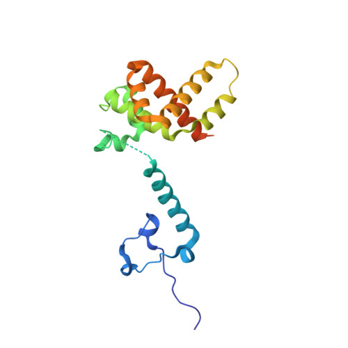
    The M2-1 protein of the important pathogen human respiratory syncytial virus is a zinc-binding transcription antiterminator that is essential for viral gene expression. We present the crystal structure of full-length M2-1 protein in its native tetrameric form at a resolution of 2.5 Å. The structure reveals that M2-1 forms a disk-like assembly with tetramerization driven by a long helix forming a four-helix bundle at its center, further stabilized by contact between the zinc-binding domain and adjacent protomers. The tetramerization helix is linked to a core domain responsible for RNA binding activity by a flexible region on which lie two functionally critical serine residues that are phosphorylated during infection. The crystal structure of a phosphomimetic M2-1 variant revealed altered charge density surrounding this flexible region although its position was unaffected. Structure-guided mutagenesis identified residues that contributed to RNA binding and antitermination activity, revealing a strong correlation between these two activities, and further defining the role of phosphorylation in M2-1 antitermination activity. The data we present here identify surfaces critical for M2-1 function that may be targeted by antiviral compounds.


  • Organizational Affiliation

    Astbury Centre for Structural Molecular Biology and School of Molecular and Cellular Biology, University of Leeds, Leeds LS2 9JT, United Kingdom.


Macromolecules
Find similar proteins by:  (by identity cutoff)  |  3D Structure
Entity ID: 1
MoleculeChains Sequence LengthOrganismDetailsImage
MATRIX PROTEIN 2-1
A, B, C, D, E
A, B, C, D, E, F, G, H, I, J, K, L, M, N, O, P
199Human respiratory syncytial virus A2Mutation(s): 0 
UniProt
Find proteins for P04545 (Human respiratory syncytial virus A (strain A2))
Explore P04545 
Go to UniProtKB:  P04545
Entity Groups  
Sequence Clusters30% Identity50% Identity70% Identity90% Identity95% Identity100% Identity
UniProt GroupP04545
Sequence Annotations
Expand
  • Reference Sequence
Small Molecules
Ligands 1 Unique
IDChains Name / Formula / InChI Key2D Diagram3D Interactions
ZN
Query on ZN

Download Ideal Coordinates CCD File 
AA [auth K]
BA [auth L]
CA [auth M]
DA [auth N]
EA [auth O]
AA [auth K],
BA [auth L],
CA [auth M],
DA [auth N],
EA [auth O],
FA [auth P],
Q [auth A],
R [auth B],
S [auth C],
T [auth D],
U [auth E],
V [auth F],
W [auth G],
X [auth H],
Y [auth I],
Z [auth J]
ZINC ION
Zn
PTFCDOFLOPIGGS-UHFFFAOYSA-N
Experimental Data & Validation

Experimental Data

  • Method: X-RAY DIFFRACTION
  • Resolution: 2.95 Å
  • R-Value Free: 0.255 
  • R-Value Work: 0.205 
  • R-Value Observed: 0.208 
  • Space Group: P 1 21 1
Unit Cell:
Length ( Å )Angle ( ˚ )
a = 91.853α = 90
b = 141.616β = 96.14
c = 141.694γ = 90
Software Package:
Software NamePurpose
REFMACrefinement
XDSdata reduction
Aimlessdata scaling
PHASERphasing

Structure Validation

View Full Validation Report


Entry History 

Deposition Data

Revision History  (Full details and data files)

  • Version 1.0: 2014-01-22
    Type: Initial release
  • Version 1.1: 2014-01-29
    Changes: Database references
  • Version 1.2: 2014-02-12
    Changes: Database references