3VTF

Structure of a UDP-glucose dehydrogenase from the hyperthermophilic archaeon Pyrobaculum islandicum


Experimental Data Snapshot

  • Method: X-RAY DIFFRACTION
  • Resolution: 2.00 Å
  • R-Value Free: 0.216 
  • R-Value Work: 0.177 
  • R-Value Observed: 0.179 

wwPDB Validation   3D Report Full Report


Ligand Structure Quality Assessment 


This is version 1.2 of the entry. See complete history


Literature

Structure of a UDP-glucose dehydrogenase from the hyperthermophilic archaeon Pyrobaculum islandicum.

Sakuraba, H.Kawai, T.Yoneda, K.Ohshima, T.

(2012) Acta Crystallogr Sect F Struct Biol Cryst Commun 68: 1003-1007

  • DOI: https://doi.org/10.1107/S1744309112030667
  • Primary Citation of Related Structures:  
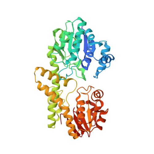
    3VTF

  • PubMed Abstract: 

    The crystal structure of an extremely thermostable UDP-glucose dehydrogenase (UDP-GDH) from the hyperthermophilic archaeon Pyrobaculum islandicum was determined at a resolution of 2.0 Å. The overall fold was comprised of an N-terminal NAD(+) dinucleotide binding domain and a C-terminal UDP-sugar binding domain connected by a long α-helix, and the main-chain coordinates of the enzyme were similar to those of previously studied UDP-GDHs, including the enzymes from Burkholderia cepacia, Streptococcus pyogenes and Klebsiella pneumoniae. However, the sizes of several surface loops in P. islandicum UDP-GDH were much smaller than the corresponding loops in B. cepacia UDP-GDH but were comparable to those of the S. pyogenes and K. pneumoniae enzymes. Structural comparison revealed that the presence of extensive intersubunit hydrophobic interactions, as well as the formation of an intersubunit aromatic pair network, is likely to be the main factor contributing to the hyperthermostability of P. islandicum UDP-GDH.


  • Organizational Affiliation

    Department of Applied Biological Science, Faculty of Agriculture, Kagawa University, 2393 Ikenobe, Kita-gun, Kagawa 761-0795, Japan.


Macromolecules
Find similar proteins by:  (by identity cutoff)  |  3D Structure
Entity ID: 1
MoleculeChains Sequence LengthOrganismDetailsImage
UDP-glucose 6-dehydrogenase444Pyrobaculum islandicum DSM 4184Mutation(s): 0 
Gene Names: Pisl_1505
EC: 1.1.1.22
UniProt
Find proteins for A1RUM9 (Pyrobaculum islandicum (strain DSM 4184 / JCM 9189 / GEO3))
Explore A1RUM9 
Go to UniProtKB:  A1RUM9
Entity Groups  
Sequence Clusters30% Identity50% Identity70% Identity90% Identity95% Identity100% Identity
UniProt GroupA1RUM9
Sequence Annotations
Expand
  • Reference Sequence
Small Molecules
Ligands 2 Unique
IDChains Name / Formula / InChI Key2D Diagram3D Interactions
UPG
Query on UPG

Download Ideal Coordinates CCD File 
B [auth A]URIDINE-5'-DIPHOSPHATE-GLUCOSE
C15 H24 N2 O17 P2
HSCJRCZFDFQWRP-JZMIEXBBSA-N
EDO
Query on EDO

Download Ideal Coordinates CCD File 
C [auth A]1,2-ETHANEDIOL
C2 H6 O2
LYCAIKOWRPUZTN-UHFFFAOYSA-N
Experimental Data & Validation

Experimental Data

  • Method: X-RAY DIFFRACTION
  • Resolution: 2.00 Å
  • R-Value Free: 0.216 
  • R-Value Work: 0.177 
  • R-Value Observed: 0.179 
  • Space Group: C 1 2 1
Unit Cell:
Length ( Å )Angle ( ˚ )
a = 117.652α = 90
b = 76.739β = 125.8
c = 75.551γ = 90
Software Package:
Software NamePurpose
DENZOdata reduction
SCALEPACKdata scaling
MOLREPphasing
REFMACrefinement
PDB_EXTRACTdata extraction
HKL-2000data collection
HKL-2000data reduction
HKL-2000data scaling

Structure Validation

View Full Validation Report


Ligand Structure Quality Assessment 


Entry History 

Deposition Data

Revision History  (Full details and data files)

  • Version 1.0: 2012-09-19
    Type: Initial release
  • Version 1.1: 2017-11-22
    Changes: Refinement description
  • Version 1.2: 2023-11-08
    Changes: Data collection, Database references, Derived calculations, Refinement description