Structural and functional conservation of key domains in InsP3 and ryanodine receptors.
Seo, M.D., Velamakanni, S., Ishiyama, N., Stathopulos, P.B., Rossi, A.M., Khan, S.A., Dale, P., Li, C., Ames, J.B., Ikura, M., Taylor, C.W.(2012) Nature 483: 108-112
- PubMed: 22286060
- DOI: https://doi.org/10.1038/nature10751
- Primary Citation of Related Structures:
3UJ0, 3UJ4 - PubMed Abstract:
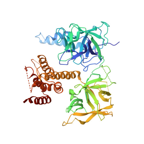
Inositol-1,4,5-trisphosphate receptors (InsP(3)Rs) and ryanodine receptors (RyRs) are tetrameric intracellular Ca(2+) channels. In each of these receptor families, the pore, which is formed by carboxy-terminal transmembrane domains, is regulated by signals that are detected by large cytosolic structures. InsP(3)R gating is initiated by InsP(3) binding to the InsP(3)-binding core (IBC, residues 224-604 of InsP(3)R1) and it requires the suppressor domain (SD, residues 1-223 of InsP(3)R1). Here we present structures of the amino-terminal region (NT, residues 1-604) of rat InsP(3)R1 with (3.6 Å) and without (3.0 Å) InsP(3) bound. The arrangement of the three NT domains, SD, IBC-β and IBC-α, identifies two discrete interfaces (α and β) between the IBC and SD. Similar interfaces occur between equivalent domains (A, B and C) in RyR1 (ref. 9). The orientations of the three domains when docked into a tetrameric structure of InsP(3)R and of the ABC domains docked into RyR are remarkably similar. The importance of the α-interface for activation of InsP(3)R and RyR is confirmed by mutagenesis and, for RyR, by disease-causing mutations. Binding of InsP(3) causes partial closure of the clam-like IBC, disrupting the β-interface and pulling the SD towards the IBC. This reorients an exposed SD loop ('hotspot' (HS) loop) that is essential for InsP(3)R activation. The loop is conserved in RyR and includes mutations that are associated with malignant hyperthermia and central core disease. The HS loop interacts with an adjacent NT, suggesting that activation re-arranges inter-subunit interactions. The A domain of RyR functionally replaced the SD in full-length InsP(3)R, and an InsP(3)R in which its C-terminal transmembrane region was replaced by that from RyR1 was gated by InsP(3) and blocked by ryanodine. Activation mechanisms are conserved between InsP(3)R and RyR. Allosteric modulation of two similar domain interfaces within an N-terminal subunit reorients the first domain (SD or A domain), allowing it, through interactions of the second domain of an adjacent subunit (IBC-β or B domain), to gate the pore.
- Ontario Cancer Institute and Department of Medical Biophysics, University of Toronto, Ontario M5G 1L7, Canada.
Organizational Affiliation: