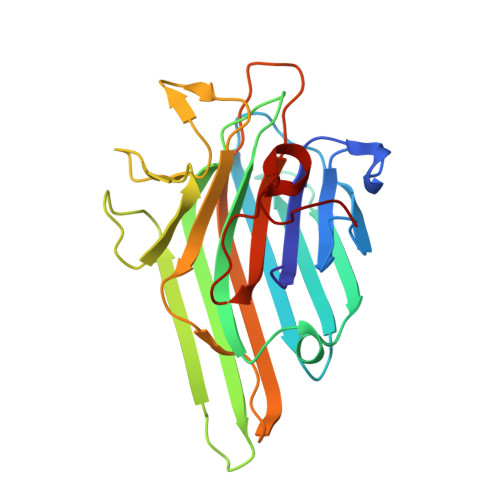
Crystal structure of the lectin of Camptosema pedicellatum: implications of a conservative substitution at the hydrophobic subsite.
Souza Teixeira, C., Colares da Silva, H., Rocha de Moura, T., Pereira-Junior, F.N., Santiago do Nascimento, K., Shiniti Nagano, C., Holanda Sampaio, A., Delatorre, P., Matias Rocha, B.A., Sousa Cavada, B.(2012) J Biochem 152: 87-98
- PubMed: 22554687
- DOI: https://doi.org/10.1093/jb/mvs047
- Primary Citation of Related Structures:
3U4X - PubMed Abstract:
Lectins have been used as models for studies of the molecular basis of protein-carbohydrate interaction and specificity by deciphering codes present in the glycan structures. The purpose of the present study was to purify and solve the complete primary and crystal structure of the lectin of Camptosema pedicellatum (CPL) complexed with 5-bromo-4-chloro-3-indolyl-α-d-mannose (X-Man) using tandem mass spectrometry. CPL was purified by single-step affinity chromatography. Mass spectrometry findings revealed that purified CPL features a combination of chains weighing 25,298 ± 2 (α-chain), 12,835 ± 2 (β-chain) and 12,481 ± 2 Da (γ-chain). The solved crystal structure of CPL features a conservative mutation in the hydrophobic subsite, a constituent of the carbohydrate recognition domain (CRD), indicating the relevance of hydrophobic interactions in the establishment of interactions with carbohydrates. The substitution and the analysis of the interactions with X-Man also revealed that the hydrophobic effect caused by a minor change in the hydrophobic subsite interferes in the formation of H-bonds due to the reorientation of the indolyl group in the CRD.
- BioMol-Lab, Laboratório de Moléculas Biologicamente Ativas, Departamento de Bioquímica e Biologia Molecular, Universidade Federal do Ceará, Fortaleza, Ceará 60440-970, Brazil.
Organizational Affiliation: