


Crystal structure of the CPH domain of the E3 ubiquitin-protein ligase HECTD1.
Walker, J.R., Qiu, L., Li, Y., Bountra, C., Wolkstrom, M., Arrowsmith, C.H., Edwards, A.M., Bochkarev, A., Dhe-Paganon, S.To be published.
Experimental Data Snapshot
Starting Model: experimental
View more details
wwPDB Validation 3D Report Full Report

Entity ID: 1 | |||||
|---|---|---|---|---|---|
| Molecule | Chains | Sequence Length | Organism | Details | Image |
| E3 ubiquitin-protein ligase HECTD1 | 89 | Homo sapiens | Mutation(s): 1 Gene Names: HECTD1, KIAA1131 EC: 6.3.2 (PDB Primary Data), 2.3.2.26 (UniProt) | 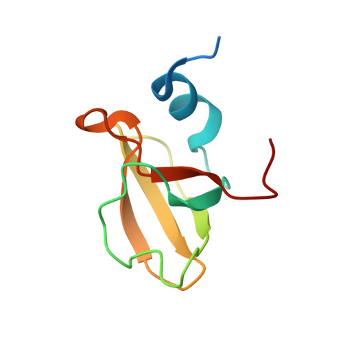 | |
UniProt & NIH Common Fund Data Resources | |||||
Find proteins for Q9ULT8 (Homo sapiens) Explore Q9ULT8 Go to UniProtKB: Q9ULT8 | |||||
PHAROS: Q9ULT8 GTEx: ENSG00000092148 | |||||
Entity Groups | |||||
| Sequence Clusters | 30% Identity50% Identity70% Identity90% Identity95% Identity100% Identity | ||||
| UniProt Group | Q9ULT8 | ||||
Sequence AnnotationsExpand | |||||
| |||||
| Length ( Å ) | Angle ( ˚ ) |
|---|---|
| a = 33.71 | α = 90 |
| b = 45.564 | β = 90 |
| c = 51.101 | γ = 90 |
| Software Name | Purpose |
|---|---|
| PHASER | phasing |
| REFMAC | refinement |
| SBC-Collect | data collection |
| HKL-2000 | data reduction |
| HKL-2000 | data scaling |


