3A2A

The structure of the carboxyl-terminal domain of the human voltage-gated proton channel Hv1


Experimental Data Snapshot

  • Method: X-RAY DIFFRACTION
  • Resolution: 2.00 Å
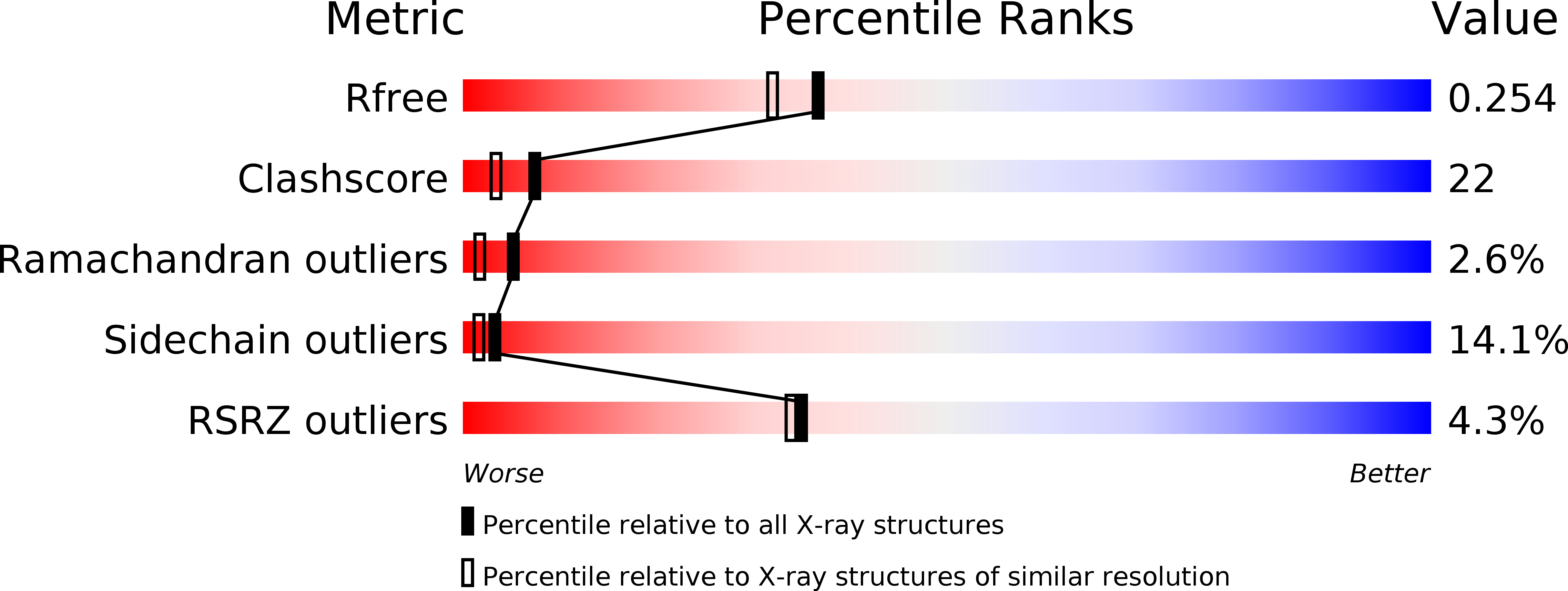
  • R-Value Free: 0.266 
  • R-Value Work: 0.252 
  • R-Value Observed: 0.253 

wwPDB Validation   3D Report Full Report


This is version 1.1 of the entry. See complete history


Literature

The role and structure of the carboxyl-terminal domain of the human voltage-gated proton channel Hv1.

Li, S.J.Zhao, Q.Zhou, Q.Unno, H.Zhai, Y.Sun, F.

(2010) J Biol Chem 285: 12047-12054

  • DOI: https://doi.org/10.1074/jbc.M109.040360
  • Primary Citation of Related Structures:  
    3A2A

  • PubMed Abstract: 

    The voltage-gated proton channel Hv1 has a voltage sensor domain but lacks a pore domain. Although the C-terminal domain of Hv1 is known to be responsible for dimeric architecture of the channel, its role and structure are not known. We report that the full-length Hv1 is mainly localized in intracellular compartment membranes rather than the plasma membrane. Truncation of either the N or C terminus alone or both together revealed that the N-terminal deletion did not alter localization, but deletion of the C terminus either alone or together with the N terminus resulted in expression throughout the cell. These results indicate that the C terminus is essential for Hv1 localization but not the N terminus. In the 2.0 A structure of the C-terminal domain, the two monomers form a dimer via a parallel alpha-helical coiled-coil, in which one chloride ion binds with the Neta atom of Arg(264). A pH-dependent structural change of the protein has been observed, but it remains a dimer irrespective of pH value.


  • Organizational Affiliation

    Key Laboratory of Bioactive Materials, Ministry of Education, College of Physics Science, Nankai University, Tianjin 300071, China. shujieli@nankai.edu.cn


Macromolecules
Find similar proteins by:  (by identity cutoff)  |  3D Structure
Entity ID: 1
MoleculeChains Sequence LengthOrganismDetailsImage
Voltage-gated hydrogen channel 1
A, B, C, D
58Homo sapiensMutation(s): 0 
UniProt & NIH Common Fund Data Resources
Find proteins for Q96D96 (Homo sapiens)
Explore Q96D96 
Go to UniProtKB:  Q96D96
PHAROS:  Q96D96
GTEx:  ENSG00000122986 
Entity Groups  
Sequence Clusters30% Identity50% Identity70% Identity90% Identity95% Identity100% Identity
UniProt GroupQ96D96
Sequence Annotations
Expand
  • Reference Sequence
Experimental Data & Validation

Experimental Data

  • Method: X-RAY DIFFRACTION
  • Resolution: 2.00 Å
  • R-Value Free: 0.266 
  • R-Value Work: 0.252 
  • R-Value Observed: 0.253 
  • Space Group: P 43
Unit Cell:
Length ( Å )Angle ( ˚ )
a = 37.65α = 90
b = 37.65β = 90
c = 137.04γ = 90
Software Package:
Software NamePurpose
REFMACrefinement
ADSCdata collection
MOSFLMdata reduction
SCALAdata scaling
SOLVEphasing

Structure Validation

View Full Validation Report


Entry History 

Deposition Data

Revision History  (Full details and data files)

  • Version 1.0: 2010-02-09
    Type: Initial release
  • Version 1.1: 2011-07-13
    Changes: Version format compliance