Activation of ATP Binding for the Autophosphorylation of Doss, a Mycobacterium Tuberculosis Histidine Kinase Lacking an ATP-Lid Motif.
Cho, H.Y., Lee, Y.H., Bae, Y.S., Kim, E., Kang, B.S.(2013) J Biological Chem 288: 12437
- PubMed: 23486471
- DOI: https://doi.org/10.1074/jbc.M112.442467
- Primary Citation of Related Structures:
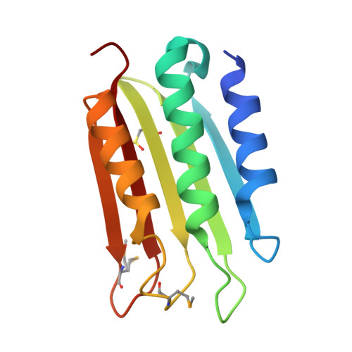
3ZXO, 3ZXQ - PubMed Abstract:
The sensor histidine kinases of Mycobacterium tuberculosis, DosS and DosT, are responsible for sensing hypoxic conditions and consist of sensor and kinase cores responsible for accepting signals and phosphorylation activity, respectively. The kinase core contains a dimerization and histidine phosphate-accepting (DHp) domain and an ATP binding domain (ABD). The 13 histidine kinase genes of M. tuberculosis can be grouped based on the presence or absence of the ATP lid motif and F box (elements known to play roles in ATP binding) in their ABDs; DosS and DosT have ABDs lacking both these elements, and the crystal structures of their ABDs indicated that they were unsuitable for ATP binding, as a short loop covers the putative ATP binding site. Although the ABD alone cannot bind ATP, the kinase core is functional in autophosphorylation. Appropriate spatial arrangement of the ABD and DHp domain within the kinase core is required for both autophosphorylation and ATP binding. An ionic interaction between Arg(440) in the DHp domain and Glu(537) in the short loop of the ABD is available and may open the ATP binding site, by repositioning the short loop away from the site. Mutations at Arg(440) and Glu(537) reduce autophosphorylation activity. Unlike other histidine kinases containing an ATP lid, which protects bound ATP, DosS is unable to accept ATP until the ABD is properly positioned relative to the histidine; this may prevent unexpected ATP reactions. ATP binding can, therefore, function as a control mechanism for histidine kinase activity.
- School of Life Science and Biotechnology, Kyungpook National University, Daegu 702-701, South Korea.
Organizational Affiliation: