3UEM

Crystal structure of human PDI bb'a' domains


Experimental Data Snapshot

  • Method: X-RAY DIFFRACTION
  • Resolution: 2.29 Å
  • R-Value Free: 0.280 
  • R-Value Work: 0.223 
  • R-Value Observed: 0.226 

wwPDB Validation   3D Report Full Report


Ligand Structure Quality Assessment 


This is version 1.2 of the entry. See complete history


Literature

Human protein-disulfide isomerase is a redox-regulated chaperone activated by oxidation of domain a'

Wang, C.Yu, J.Huo, L.Wang, L.Feng, W.Wang, C.-C.

(2012) J Biol Chem 287: 1139-1149

  • DOI: https://doi.org/10.1074/jbc.M111.303149
  • Primary Citation of Related Structures:  
    3UEM

  • PubMed Abstract: 

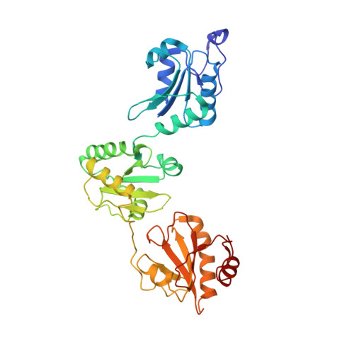
    Protein-disulfide isomerase (PDI), with domains arranged as abb'xa'c, is a key enzyme and chaperone localized in the endoplasmic reticulum (ER) catalyzing oxidative folding and preventing misfolding/aggregation of proteins. It has been controversial whether the chaperone activity of PDI is redox-regulated, and the molecular basis is unclear. Here, we show that both the chaperone activity and the overall conformation of human PDI are redox-regulated. We further demonstrate that the conformational changes are triggered by the active site of domain a', and the minimum redox-regulated cassette is located in b'xa'. The structure of the reduced bb'xa' reveals for the first time that domain a' packs tightly with both domain b' and linker x to form one compact structural module. Oxidation of domain a' releases the compact conformation and exposes the shielded hydrophobic areas to facilitate its high chaperone activity. Thus, the study unequivocally provides mechanistic insights into the redox-regulated chaperone activity of human PDI.


  • Organizational Affiliation

    National Laboratory of Biomacromolecules, Institute of Biophysics, Chinese Academy of Sciences, Beijing 100101, China.


Macromolecules
Find similar proteins by:  (by identity cutoff)  |  3D Structure
Entity ID: 1
MoleculeChains Sequence LengthOrganismDetailsImage
Protein disulfide-isomerase361Homo sapiensMutation(s): 0 
Gene Names: PDI
EC: 5.3.4.1
UniProt & NIH Common Fund Data Resources
Find proteins for P07237 (Homo sapiens)
Explore P07237 
Go to UniProtKB:  P07237
PHAROS:  P07237
GTEx:  ENSG00000185624 
Entity Groups  
Sequence Clusters30% Identity50% Identity70% Identity90% Identity95% Identity100% Identity
UniProt GroupP07237
Sequence Annotations
Expand
  • Reference Sequence
Small Molecules
Ligands 1 Unique
IDChains Name / Formula / InChI Key2D Diagram3D Interactions
D1D
Query on D1D

Download Ideal Coordinates CCD File 
B [auth A](4S,5S)-1,2-DITHIANE-4,5-DIOL
C4 H8 O2 S2
YPGMOWHXEQDBBV-QWWZWVQMSA-N
Experimental Data & Validation

Experimental Data

  • Method: X-RAY DIFFRACTION
  • Resolution: 2.29 Å
  • R-Value Free: 0.280 
  • R-Value Work: 0.223 
  • R-Value Observed: 0.226 
  • Space Group: P 21 21 21
Unit Cell:
Length ( Å )Angle ( ˚ )
a = 63.585α = 90
b = 76.076β = 90
c = 79.656γ = 90
Software Package:
Software NamePurpose
HKL-2000data collection
PHASERphasing
REFMACrefinement
HKL-2000data reduction
HKL-2000data scaling

Structure Validation

View Full Validation Report


Ligand Structure Quality Assessment 


Entry History 

Deposition Data

Revision History  (Full details and data files)

  • Version 1.0: 2011-11-23
    Type: Initial release
  • Version 1.1: 2013-06-26
    Changes: Database references
  • Version 1.2: 2024-03-20
    Changes: Data collection, Database references, Derived calculations