Replacement of the axial copper ligand methionine with lysine in amicyanin converts it to a zinc-binding protein that no longer binds copper.
Sukumar, N., Choi, M., Davidson, V.L.(2011) J Inorg Biochem 105: 1638-1644
- PubMed: 22071089
- DOI: https://doi.org/10.1016/j.jinorgbio.2011.08.002
- Primary Citation of Related Structures:
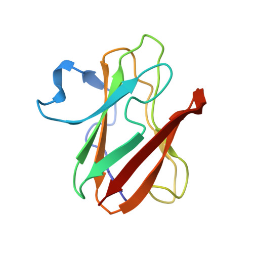
3RYM - PubMed Abstract:
The mutation of the axial ligand of the type I copper protein amicyanin from Met to Lys results in a protein that is spectroscopically invisible and redox inactive. M98K amicyanin acts as a competitive inhibitor in the reaction of native amicyanin with methylamine dehydrogenase indicating that the M98K mutation has not affected the affinity for its natural electron donor. The crystal structure of M98K amicyanin reveals that its overall structure is very similar to native amicyanin but that the type I binding site is occupied by zinc. Anomalous difference Fourier maps calculated using the data collected around the absorption edges of copper and zinc confirm the presence of Zn(2+) at the type I site. The Lys98 NZ donates a hydrogen bond to a well-ordered water molecule at the type I site which enhances the ability of Lys98 to provide a ligand for Zn(2+). Attempts to reconstitute M98K apoamicyanin with copper resulted in precipitation of the protein. The fact that the M98K mutation generated such a selective zinc-binding protein was surprising as ligation of zinc by Lys is rare and this ligand set is unique for zinc.
- NE-CAT and Department of Chemistry and Chemical Biology, Cornell University, Argonne National Laboratory, Argonne, IL 60439, USA. sukumar@anl.gov
Organizational Affiliation: