3P87

Structure of human PCNA bound to RNASEH2B PIP box peptide


Experimental Data Snapshot

  • Method: X-RAY DIFFRACTION
  • Resolution: 2.99 Å
  • R-Value Free: 0.248 
  • R-Value Work: 0.223 
  • R-Value Observed: 0.224 

wwPDB Validation   3D Report Full Report


This is version 1.3 of the entry. See complete history


Literature

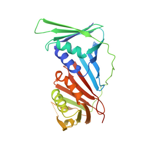
PCNA directs type 2 RNase H activity on DNA replication and repair substrates.

Bubeck, D.Reijns, M.A.Graham, S.C.Astell, K.R.Jones, E.Y.Jackson, A.P.

(2011) Nucleic Acids Res 39: 3652-3666

  • DOI: https://doi.org/10.1093/nar/gkq980
  • Primary Citation of Related Structures:  
    3P83, 3P87

  • PubMed Abstract: 

    Ribonuclease H2 is the major nuclear enzyme degrading cellular RNA/DNA hybrids in eukaryotes and the sole nuclease known to be able to hydrolyze ribonucleotides misincorporated during genomic replication. Mutation in RNASEH2 causes Aicardi-Goutières syndrome, an auto-inflammatory disorder that may arise from nucleic acid byproducts generated during DNA replication. Here, we report the crystal structures of Archaeoglobus fulgidus RNase HII in complex with PCNA, and human PCNA bound to a C-terminal peptide of RNASEH2B. In the archaeal structure, three binding modes are observed as the enzyme rotates about a flexible hinge while anchored to PCNA by its PIP-box motif. PCNA binding promotes RNase HII activity in a hinge-dependent manner. It enhances both cleavage of ribonucleotides misincorporated in DNA duplexes, and the comprehensive hydrolysis of RNA primers formed during Okazaki fragment maturation. In addition, PCNA imposes strand specificity on enzyme function, and by localizing RNase H2 and not RNase H1 to nuclear replication foci in vivo it ensures that RNase H2 is the dominant RNase H activity during nuclear replication. Our findings provide insights into how type 2 RNase H activity is directed during genome replication and repair, and suggest a mechanism by which RNase H2 may suppress generation of immunostimulatory nucleic acids.


  • Organizational Affiliation

    Division of Structural Biology, Wellcome Trust Centre for Human Genetics, University of Oxford, Oxford OX3 7BN, UK.


Macromolecules
Find similar proteins by:  (by identity cutoff)  |  3D Structure
Entity ID: 1
MoleculeChains Sequence LengthOrganismDetailsImage
Proliferating cell nuclear antigen261Homo sapiensMutation(s): 0 
Gene Names: PCNA
UniProt & NIH Common Fund Data Resources
Find proteins for P12004 (Homo sapiens)
Explore P12004 
Go to UniProtKB:  P12004
PHAROS:  P12004
GTEx:  ENSG00000132646 
Entity Groups  
Sequence Clusters30% Identity50% Identity70% Identity90% Identity95% Identity100% Identity
UniProt GroupP12004
Sequence Annotations
Expand
  • Reference Sequence

Find similar proteins by:  Sequence   |   3D Structure  

Entity ID: 2
MoleculeChains Sequence LengthOrganismDetailsImage
Ribonuclease H2 subunit B23Homo sapiensMutation(s): 0 
UniProt & NIH Common Fund Data Resources
Find proteins for Q5TBB1 (Homo sapiens)
Explore Q5TBB1 
Go to UniProtKB:  Q5TBB1
PHAROS:  Q5TBB1
GTEx:  ENSG00000136104 
Entity Groups  
Sequence Clusters30% Identity50% Identity70% Identity90% Identity95% Identity100% Identity
UniProt GroupQ5TBB1
Sequence Annotations
Expand
  • Reference Sequence
Experimental Data & Validation

Experimental Data

  • Method: X-RAY DIFFRACTION
  • Resolution: 2.99 Å
  • R-Value Free: 0.248 
  • R-Value Work: 0.223 
  • R-Value Observed: 0.224 
  • Space Group: P 1 21 1
Unit Cell:
Length ( Å )Angle ( ˚ )
a = 83.108α = 90
b = 81.788β = 91.47
c = 116.558γ = 90
Software Package:
Software NamePurpose
ADSCdata collection
PHASERphasing
BUSTERrefinement
MOSFLMdata reduction
SCALAdata scaling

Structure Validation

View Full Validation Report


Entry History 

Deposition Data

Revision History  (Full details and data files)

  • Version 1.0: 2011-02-02
    Type: Initial release
  • Version 1.1: 2011-07-13
    Changes: Version format compliance
  • Version 1.2: 2014-04-02
    Changes: Structure summary
  • Version 1.3: 2023-09-06
    Changes: Data collection, Database references, Refinement description