3NH7

Crystal structure of the neutralizing Fab fragment AbD1556 bound to the BMP type I receptor IA


Experimental Data Snapshot

  • Method: X-RAY DIFFRACTION
  • Resolution: 2.70 Å
  • R-Value Free: 0.280 
  • R-Value Work: 0.234 
  • R-Value Observed: 0.236 

wwPDB Validation   3D Report Full Report


This is version 1.2 of the entry. See complete history


Literature

A selection fit mechanism in BMP receptor IA as a possible source for BMP ligand-receptor promiscuity

Harth, S.Kotzsch, A.Hu, J.Sebald, W.Mueller, T.D.

(2010) PLoS One 5: e13049-e13049

  • DOI: https://doi.org/10.1371/journal.pone.0013049
  • Primary Citation of Related Structures:  
    3NH7

  • PubMed Abstract: 

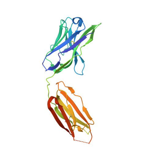
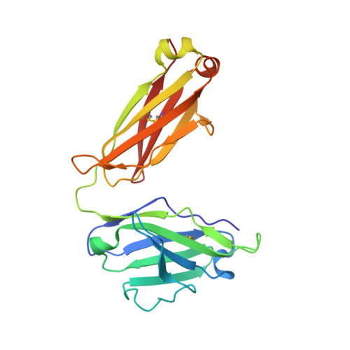
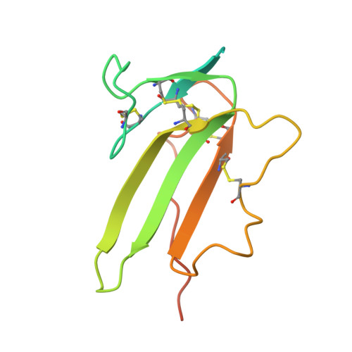
    Members of the TGF-β superfamily are characterized by a highly promiscuous ligand-receptor interaction as is readily apparent from the numeral discrepancy of only seven type I and five type II receptors available for more than 40 ligands. Structural and functional studies have been used to address the question of how specific signals can be deduced from a limited number of receptor combinations and to unravel the molecular mechanisms underlying the protein-protein recognition that allow such limited specificity. In this study we have investigated how an antigen binding antibody fragment (Fab) raised against the extracellular domain of the BMP receptor type IA (BMPR-IA) recognizes the receptor's BMP-2 binding epitope and thereby neutralizes BMP-2 receptor activation. The crystal structure of the complex of the BMPR-IA ectodomain bound to the Fab AbD1556 revealed that the contact surface of BMPR-IA overlaps extensively with the contact surface for BMP-2 interaction. Although the structural epitopes of BMPR-IA to both binding partners coincides, the structures of BMPR-IA in the two complexes differ significantly. In contrast to the structural differences, alanine-scanning mutagenesis of BMPR-IA showed that the functional determinants for binding to the antibody and BMP-2 are almost identical. Comparing the structures of BMPR-IA bound to BMP-2 or bound to the Fab AbD1556 with the structure of unbound BMPR-IA shows that binding of BMPR-IA to its interaction partners follows a selection fit mechanism, possibly indicating that the ligand promiscuity of BMPR-IA is inherently encoded by structural adaptability. The functional and structural analysis of the BMPR-IA binding antibody AbD1556 mimicking the BMP-2 binding epitope may thus pave the way for the design of low-molecular weight synthetic receptor binders/inhibitors.


  • Organizational Affiliation

    Lehrstuhl für Physiologische Chemie II, Theodor-Boveri-Institut für Biowissenschaften der Universität Würzburg, Würzburg, Germany.


Macromolecules
Find similar proteins by:  (by identity cutoff)  |  3D Structure
Entity ID: 1
MoleculeChains Sequence LengthOrganismDetailsImage
Antibody fragment Fab AbD1556, heavy chainA [auth H],
D [auth I],
G [auth J],
J [auth K]
234Homo sapiensMutation(s): 0 
Entity Groups  
Sequence Clusters30% Identity50% Identity70% Identity90% Identity95% Identity100% Identity
Sequence Annotations
Expand
  • Reference Sequence
Find similar proteins by:  (by identity cutoff)  |  3D Structure
Entity ID: 2
MoleculeChains Sequence LengthOrganismDetailsImage
Antibody fragment Fab AbD1556, light chainB [auth L],
E [auth M],
H [auth N],
K [auth O]
213Homo sapiensMutation(s): 0 
Entity Groups  
Sequence Clusters30% Identity50% Identity70% Identity90% Identity95% Identity100% Identity
Sequence Annotations
Expand
  • Reference Sequence
Find similar proteins by:  (by identity cutoff)  |  3D Structure
Entity ID: 3
MoleculeChains Sequence LengthOrganismDetailsImage
Bone morphogenetic protein receptor type-1AC [auth A],
F [auth B],
I [auth C],
L [auth D]
129Homo sapiensMutation(s): 0 
Gene Names: BMPR1A
EC: 2.7.11.30
UniProt & NIH Common Fund Data Resources
Find proteins for P36894 (Homo sapiens)
Explore P36894 
Go to UniProtKB:  P36894
PHAROS:  P36894
GTEx:  ENSG00000107779 
Entity Groups  
Sequence Clusters30% Identity50% Identity70% Identity90% Identity95% Identity100% Identity
UniProt GroupP36894
Sequence Annotations
Expand
  • Reference Sequence
Experimental Data & Validation

Experimental Data

  • Method: X-RAY DIFFRACTION
  • Resolution: 2.70 Å
  • R-Value Free: 0.280 
  • R-Value Work: 0.234 
  • R-Value Observed: 0.236 
  • Space Group: P 1 21 1
Unit Cell:
Length ( Å )Angle ( ˚ )
a = 89.32α = 90
b = 129.255β = 92.27
c = 100.239γ = 90
Software Package:
Software NamePurpose
CrystalCleardata collection
PHASERphasing
REFMACrefinement
CrystalCleardata reduction
CrystalCleardata scaling

Structure Validation

View Full Validation Report


Entry History 

Deposition Data

Revision History  (Full details and data files)

  • Version 1.0: 2010-10-27
    Type: Initial release
  • Version 1.1: 2011-07-13
    Changes: Version format compliance
  • Version 1.2: 2023-11-01
    Changes: Data collection, Database references, Refinement description